Received 14 May 2006 | Accepted 7 September 2006 | Published 13 September 2006
Reprint
Received 14 May 2006 | Accepted 7 September 2006 | Published 13 September 2006 |
Download Reprint |
Immunochemical detection of glycated β- and γ-crystallins in lens and their circulating autoantibodies (IgG) in streptozocin induced diabetic rat
Mala Ranjan,
Sujatha Nayak,
Beedu Sashidhar Rao
Department of Biochemistry, University College of Science, Osmania University, Hyderabad, India
Correspondence to: Dr. B. Sashidhar Rao, Department of Biochemistry, University College of Science, Osmania University, Hyderabad-500 007 (A.P.) India; Phone: +91-040-27016868; FAX: +91-040-27016868; email: sashi_rao@yahoo.com
Abstract
Purpose: This study used an immunochemical approach aimed to detect the glycated crystallins (β- and γ-crystallin) in rat lens and their circulating specific autoantibodies in serum during the course of cataractogenesis.
Methods: Streptozocin (STZ; 55 mg/kg body mass) induced diabetic male Wistar/NIN rats (2-3 months old) and control nondiabetic rats were used for this study. Plasma glucose, glycated hemoglobin and body weight were evaluated on day zero, and at the interval of every two weeks up to the eighth week of post-injection in both the groups. Other biochemical parameters, such as the levels of nonprotein sulfhydryl (-SH) groups and the activity of γ-glutamyl transpeptidase (γ-GT) in lens proteins were also estimated. Cataract progress was monitored by measuring the advanced glycation end product (AGE)-like fluorophores in both intact lens as well as in lens homogenate employing digital based image analysis and spectrofluorimetric methods. Similarly, the polyclonal antibodies specific to β-glycated-, γ-glycated-, β-, and γ-crystallins were used to determine the concentration of respective immunogens in lens by noncompetitive ELISA and their respective circulating antibodies by antibody capture assay. The profile of glycated lens protein (soluble and insoluble fractions) during the course of cataractogenesis was assessed by the western blot technique.
Results: STZ induced diabetic rats showed typical signs of diabetes (hyperglycemia, increased water and food intake with no increase in body weight). Biochemical analysis of total lens protein showed a significant (p=<0.001) decrease in the levels of nonprotein -SH groups. The activity of lenticular γ-GT in diabetic rats was found to be unaltered as compared to the control group. Digital analysis of intact lens illustrated a positive correlation (r2=0.888) with the formation of AGE-like fluorophores during the course of cataractogenesis. A similar trend was also observed in the levels of AGE-like fluorophores in the total lens homogenate of diabetic animals during the course of cataractogenesis. The concentration of β- and γ-glycated-crystallins in the rat lens (soluble and insoluble fractions) was analyzed by non-competitive ELISA. The concentration of β- and γ-glycated-crystallins were found to be enhanced by the end of week eight, as compared to the control group. Concomitantly, crystallin-specific (β- and γ-glycated-crystallin) autoantibodies were also detected in the serum of the diabetic rats from week two onwards. Western blot analysis indicated the formation of enhanced glycated lens crystallins (β- and γ-crystallin) in the insoluble fraction.
Conclusions: The following was observed during the course of cataractogenesis: (1) there was an enhanced formation of AGEs-like fluorophores in intact lens; (2) β- and γ-glycated-crystallin levels increased in the rat lens (insoluble fraction) by the end of week eight; and (3) release of these glycated lens proteins into peripheral circulation resulted in the production of autoantibodies to β- and γ-glycated-crystallins that could be detected as early as week two, after induction of diabetic status in experimental rats.
Introduction
Diabetes has been identified as a key risk factor for cataract [1]. There is an uncontrolled diffusion of extracellular glucose into the lens, which is one of the important body parts affected in diabetes mellitus. The proteins in the lens are extremely long lived, and there is virtually no protein turnover, which provides great opportunity for posttranslational modification to occur. Several mechanisms have been implicated in the development of cataract in diabetes [2-4]. The role of Maillard reactions in cataract formation has also been extensively studied in both aged and diabetic lens wherein advanced glycation end products (AGEs) of various derivations and molecular structures have been shown to be markedly elevated [5-8].
Crystallins are the major structural proteins of the eye lens and account for approximately 90% of total proteins. All vertebrate lenses contain α-, β-, and γ-crystallins. Crystallins undergo nonenzymatic glycation [9] and their degree of glycation have been checked by affinity chromatography [10] as well as immunochemically using polyclonal and monoclonal antibodies to glycated bovine serum albumin (BSA-AGE) in streptozocin (STZ) induced diabetic rats [11,12]. During the course of aging, high-molecular-weight (HMW) and insoluble proteins increase with a concomitant disappearance of γ-crystallins from soluble fraction. This disappearance of γ-crystallins coincides with increased glycation and decrease in sulfhydryl (-SH) groups [13] from soluble fraction. It appears that glycation of lens proteins, disappearance of γ-crystallins, alterations in the -SH groups in the soluble fraction, increase in insoluble fraction, and HMW aggregates are interrelated. Leakage of γ-crystallins into the vitreous humor [14] is one of the first chronological events at the time of progression of opacity in diabetic rat that was observed along with the covalent modification of membrane proteins. Immunologically, it has also been observed that apart from the γ-crystallins, there are other subunits of α- and β-crystallins outside the lens [15]. It has been reported that more than 98% of the cataract patients showed elevated antibody titer against lens crystallins [16]. Few studies have suggested that age-related cataract (ARC) may involve autoimmune phenomena [17-21]. The presence of these circulating autoantibodies are found to be cytotoxic for lens epithelial cells [22]. These observations have explored the therapeutic value of oral administration of lens proteins in ARC [23]. Hence, it is pertinent to note that autoimmune phenomena could play a key role in the initiation and progression of cataract.
Anticrystallin autoantibodies have often been demonstrated in the serum of healthy persons and especially in patients with cataract [15]. In no case, however the specific lens glycated crystallins have been identified against their glycated crystallin specific antibodies during the course of cataractogenesis. Since, the lens structural proteins have a fairly high degree of organ specificity [24], their elevated concentrations in blood serum might signal impairment of normal lens physiology leading to risk of developing cataract. In order to explore the feasibility of using serum assays of lens proteins to gain information on the physiological status of the lens, we developed a noncompetitive enzyme immunoassay to detect the glycated β- and γ-crystallins in lens and their circulating autoantibodies (IgG) in STZ induced diabetic rat, during the course of cataractogenesis.
Methods
Materials
Bovine serum albumin (BSA-fatty acid free, RIA grade), Freund's complete adjuvant (FCA), Freund's incomplete adjuvant (FIA), fish gelatin, hemoglobin, antirabbit immunoglobulin G (IgG) labeled with horseradish peroxidase raised in goat (whole molecule), tetra methyl benzidine (TMB), urea hydrogen peroxide, β-cyclodextrin, antirat IgG labeled with horseradish peroxidase raised in goat, p-nitro-blue tetrazolium chloride (NBT), 5-bromo-4-chloro-3-indolyl phosphate toluidine (BCIP), STZ, Immobilon-P transfer membrane and Sepharose 6B were purchased from Sigma (St. Louis, MO). Polystyrene microtiter ELISA plates were from Greiner, Nurtingen, Germany. Other reagents used were of analytical grade.
Experimental design
Male, Wistar/NIN rats (2-3 months old) with a mean body mass of 250 g were obtained from the National Center for Laboratory Animal Sciences, National Institute of Nutrition, Hyderabad. All animals were fed on a cereals-plus based semisynthetic stock pellet diet. The control rats (Group I; n=30) received citrate buffer (0.05 M, pH 4.5) as a vehicle, where as the experimental rats (Group II; n=50) received a single intraperitoneal injection of STZ (55 mg/kg body mass) in the same buffer [25].
Animal care protocols were in accordance with and approved by the Institutional Animal Ethics Committee. Animals were housed in individual cages in a temperature and humidity controlled room having a 12 h light:dark cycle. Food and water were provided ad libitum. Body mass was monitored weekly.
Every two weeks (zero, two, four, six, and eight) four animals each (control and experimental) were sacrificed. Blood was drawn from the retroorbital plexus from each animal for determination of plasma glucose level, total hemoglobin, and glycated hemoglobin as indicators of hyperglycemia. Lenses were dissected by posterior approach from each animal and stored at -20 °C until further analysis.
Digital-based image analysis of rat lens
This system consisted of a high-resolution charged coupled device (CCD) based camera with absorbance and fluorescence facility. It had an optical zoom lens (12.5x75 mm/f1.8) along with a 49 mm +1 dioptres close-up lens. The sensitivity of the camera was 10-5 lux, with negligible signal to noise ratio (<30 db). The CCD camera was housed in a light-tight compact cabinet over a transilluminator and was equipped with UV and IR interference filters. The acquired image is displayed on a built in LCD screen (resolution -8 bit, 256 gray level images). Image acquisition was base on real-time integration in the range of 0.04 to 10 s.
Five lenses from both groups for every time were selected for digital-based image analysis. Digital image acquisition was achieved by placing the intact lens on the flat surface of the transilluminator, housed in the light-tight cabinet. The sample (lens) was excited individually from the base at UV-A (λex 365 nm) region of the electromagnetic spectrum. Lens image formed due to the emission at 450 nm was acquired by real-time integration at 0.04 s. Annotated images were saved in a PC compatible file format (.tif file) to a floppy disk. Later, the digital images of the intact lens were analyzed by the software for determining the net fluorescence density (due to the emitted intensity of the light) measured as peak volume. This was done to measure the AGE-like fluorophores, as indicated by fluorescent intensity (peak volume) at λex 365 nm and λem 450 nm in intact rat lens using high sensitivity CCD-based digital image analyzer (UVItec, Cambridge, UK). Fluorescent intensity (peak volume) was analyzed using UVI image acquisition and analysis software [26].
Biochemical assessment of diabetic status
Plasma glucose levels were determined by glucose oxidase-peroxidase assay method [27]. Total hemoglobin and glycosylated hemoglobin were measured by an ion exchange resin method [28]. The concentration of nonprotein -SH groups in the lens proteins was estimated spectrophotometrically by the method of Sedlack and Lindsay [29]. γ-GT activity was measure by the method of Tate and Meister [30].
Preparation of rat lens homogenates
Lenses (n=2) were homogenized individually in 0.02 M phosphate buffer (pH 7.4; 10% w/v), centrifuged at 10,000x g for 30 min at 4 °C, and separated into supernatant and precipitate. The supernatant was referred to as the "buffer soluble" fraction and precipitate as the "buffer insoluble" fraction. The precipitate was further solubilized in 200 ml of 0.1 N NaOH. Both fractions were used for ELISA and western blot analysis. Total homogenate was also analyzed to detect the level of AGE-like fluorophores [26]. Protein was estimated by the method of Lowry et al. [31] using BSA as a reference standard.
Fluorescence measurements
Fluorescent measurements were performed in total lens homogenate samples, using a spectrofluorimeter (Perkin-Elmer, LS-3B, Norwalk, NJ). AGEs were measured as described in previous research [32,33] at λex 366 nm and λem 440 nm. Results are expressed as fluorescent intensity (FI)/mg of lens protein.
Production and characterization of polyclonal antibodies to crystallins (β- and γ-crystallin) and glycated crystallins (β-gly- and γ-gly-crystallin)
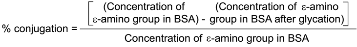
Lens β- and γ-crystallins were isolated from one-month-old male Wistar/NIN rats using Sepharose-6B column chromatography, by following a technique previously described by Bhat [34]. Glycated β- and γ-crystallins were synthesized in vitro as described elsewhere in the literature [35]. Briefly, proteins (β- and γ-crystallins; 0.1 g) were dissolved along with glucose (2.9 g) and ribose (0.1 g) in 5 ml of 0.4 M sterile sodium phosphate buffer (pH 7.4). Samples were processed under sterile condition using the laminar-flow cabinet. Vials were sealed and placed in an air-circulating incubator at 37 °C for 12 weeks. After incubation, the mixture was dialyzed extensively against 0.02 M sodium phosphate buffer (pH 7.4) containing 0.15 M NaCl. The degree of glycation was assessed by the trinitobenzene sulfonic acid method [36].
Polyclonal antibodies for crystallins (β- and γ-crystallin) and glycated crystallins (β-gly- and γ-gly-crystallin) were raised in rabbits following guidelines in the literature [32]. A primer dose of 250-300 μg/kg of body mass of the purified crystallins (β- and γ-crystallin) and synthesized glycated crystallins (β-gly- and γ-gly-crystallin) were injected at multiple sites (intradermal injection). It was dissolved in sterile saline and emulsified with Freund's complete adjuvant in a 1:1 ratio. Later, three booster doses were given (100-150 μg/kg) intramuscularly in Freund's incomplete adjuvant. Blood was collected from the animals after 30 days of primer dose and 10-12 days after the booster doses. At the end of the immunization schedule the animals were sacrificed and blood was collected by cardiac puncture. Serum was separated, lyophilized, and stored at -20 °C until further use. Immune response to these immunogens was checked by the Ouchterlony double diffusion technique [37]. Antisera titer for preimmune, primer, I, II, and III boosters were determined by checkerboard analysis using antibody capture assay. The cross-reactivity of these antisera was checked against glycated-hemoglobin.
Noncompetitive ELISA
The polyclonal antibodies specific to β-, γ-, β-gly- and γ-gly-crystallins were used to determine the levels of respective immunogens in lens by noncompetitive ELISA as described in detail previously with minor modifications [38]. Briefly, a 96 well microtiter plate was coated with a concentration of antigens (β-, γ- crystallins, 500 ng/50 μl/well and β-gly-, γ-gly-crystallins, 100 ng/50 μl/well) dissolved in 0.1 M carbonate buffer, pH 9.6, overnight at 37 °C and washed thrice with washing buffer (phosphate buffer saline [PBS-T] 0.01 M, pH 7.2 with 0.05% tween-20 and 0.01% sodium azide). The wells were blocked for nonspecific binding with 100 μl/well of 0.1% fish gelatin in PBS (0.01 M, pH 7.2) and washed three times as earlier. To each well was added 50 μl of diluted respective polyclonal antibodies (β-, 1:7,000; γ-, β-gly-, and γ-gly-crystallin, 1:10,000), which had been preincubated with test (lens sample) and standard at different concentrations (1-1000 ng) of their respective antigens for 1 h at room temperature prior to addition to the plate. After 1 h incubation, wells were washed thrice and later incubated at 37 °C for 1 h with 50 μl/well of 1:6,000 diluted (0.5% fish gelatin in PBS) horseradish peroxidase labeled antirabbit IgG raised in goat. The plates were washed thrice and 150 μl/well of substrate (450 μl of TMB [10 mg/ml of dimethyl sulfoxide] in 15 ml of 0.1 M acetate buffer pH 5.0 containing 0.25% (w/v) β-cyclodextrin and 0.015% (w/v) of urea hydrogen peroxide) was added. The reaction was terminated after 10 min by adding 50 μl/well of 1.25 M H2SO4. The absorbance was recorded at 450 nm using a SLT Spectra ELISA reader (SLT Lab Instruments, Salzburg, Austria). Percent binding of antibodies versus concentration of analyte (1-1000 ng) was used to generate inhibition plot using linear regression analysis. The concentration of respective antigens (β-, γ-, β-gly-, and γ-gly-crystallins) in lens was expressed as μg/mg of lens protein. Concentration was defined as the amount of proteins (β-, γ-, β-gly-, and γ-gly-crystallins) required to inhibit 50% (IC50) of their respective antibody binding.
Crossreactivity of the respective antibodies (β-, γ-, β-gly-, and γ-gly-crystallins) to β-, γ-, β-gly-, and γ-gly-crystallins were evaluated by noncompetitive ELISA wherein these antigens (β-, γ-, β-gly-, and γ-gly-crystallins) were used as analytes to displace the antibody. Cross-reactivity studies were carried out at 50% inhibition (IC50) level of the respective parent antigens.
Antibody capture assay
The concentration of circulating antibodies to β-, γ-, β-gly-, and γ-gly-crystallins in serum from experimental rats were analyzed by antibody capture assay as described previously [38] using their respective antigens (β-, γ-, β-gly-, and γ-gly-crystallins) from the same species. Microtiter plates were coated with their respective concentration of antigens (β- and γ- crystallins separately, 500 ng/50 μl/well and β-gly- and γ-gly-crystallins separately, 100 ng/50 μl/well) dissolved in 0.1 M carbonate buffer, pH 9.6, incubated overnight at 37 °C and washed thrice with washing buffer. The wells were blocked for nonspecific binding with 100 μl/well of 0.1% fish gelatin in PBS (0.01 M, pH 7.2). Plates were washed thrice, and different dilution of antisera (experimental and control) from rats were added and incubated for 2 h at 37 °C. After washing, plates were incubated at 37 °C for 1 h with 50 μl/well of 1:6,000 diluted (0.5% fish gelatin in PBS) horseradish peroxidase labeled anti-rat IgG raised in goat. The plates were washed thrice and 150 μl/well of substrate (450 μl of TMB [10 mg/ml of dimethyl sulfoxide]) in 15 ml of 0.1 M acetate buffer pH 5.0 containing 0.25% (w/v) β-cyclodextrin and 0.015% (w/v) of urea hydrogen peroxide was added. The reaction was terminated after 10 min by adding 50 μl/well of 1.25 M H2SO4. The absorbance was recorded at 450 nm using SLT Spectra ELISA reader (SLT Lab Instruments, Salzburg, Austria). Rat IgG was used to generate the calibration plot for crystallin specific antibodies (β-, γ-, β-gly-, and γ-gly-crystallins). Results were expressed as μg/ml of serum.
Western blot analysis
Western blot analysis was carried out according to the Towbin method [39] with a modification that involved using 20 μg of lens protein (soluble and insoluble fractions) of week eight from both the groups, to assess the degree of glycation. Briefly, proteins separated by 12.5% SDS-PAGE were electrophoretically transferred to Immobilon-P transfer membrane (polyvinylidene fluoride membrane) using a Consort electroblotter system (Consort N.V., Turnhout, Belgium). The membrane was soaked in 100% methanol for 30 s and washed with water for 2 min. It was then soaked in Towbin's transfer buffer (0.025 M Tris, 0.192 M glycine and 20% methanol) for 30 min. Filter papers for sandwiching the membrane and gel were cut to the dimensions of gel and were also soaked in Towbin's transfer buffer for 30 min. The transfer was carried out at 1.1 mA/cm2 of gel, 50 V, 150 W for 2 h at 21 °C. The membrane was washed thrice with washing buffer (0.01 M PBS with 0.05% Tween-20) and incubated in blocking buffer (3% fish gelatin in 0.01 M PBS) for 30 min and washed with washing buffer. It was incubated overnight with blocking buffer containing 1:1,000 diluted polyclonal antibodies to β- and γ-glycated-crystallins with gentle shaking at 21 °C. The membrane was washed thrice with washing buffer to remove unbound antibodies and incubated for 1 h in alkaline phosphatase assay buffer (0.1 M NaCl, 0.005 M MgCl2, 0.1 M Tris, pH 9.5) containing 1:5,000 goat antirabbit IgG coupled with alkaline phosphatase. The membrane was washed thrice with washing buffer and incubated with BCIP/NBT developing solution (115 mmoles of BCIP and 61 mmoles of NBT) until purple bands appeared. Color development was arrested by immersing the membrane in water. The membrane was air dried for 20 min and stored. It was documented using a digital based image analyzer (UVItec, Cambridge, England).
Statistical analysis
The data was statistically analyzed by using Sigma-plot software version 5.0. The test of significance was based on Student's T-test.
Results
Physical and biochemical assessment of diabetic model
There was no change in body mass at the end of week eight between control (299±42 g) and STZ rats (274±52 g). Table 1 depicts the plasma glucose and glycated hemoglobin levels in control and experimental animals. The plasma glucose level in the diabetic group was found to be enhanced from week two onward to a hyperglycemic state (in the range of 350-450 mg/dl), with a concomitant increase in the concentration of glycated hemoglobin (15%). In addition, a three-fold decrease in lens protein-free -SH groups was also observed on week two as compared to week zero sample, in the diabetic animals. Further, there was no alteration in the activity of lens γ-GT during cataract progression (Table 2).
Fluorometric determination of advanced glycation end product-like fluorophores (λex 366 nm, λem 440 nm) in lens
AGE-like fluorophores were analyzed in both intact lens (Figure 1) as well as in lens homogenate (Figure 2). A positive correlation (r2=0.888) was found between the progression of cataract and fluorescent intensity in the diabetic lens, although this was not observed in the control group (Figure 1). Similarly, AGE-like fluorophores showed a statistically significant (p=<0.001) increase in diabetic lens homogenate from week two onwards, as compared to the control group (Figure 2).
Trinitrobenzene sulfonate assay
The degree of glycation of crystallins (β- and γ-crystallin) was monitored by trinitrobenzene sulfonate (TNBS) assay and was observed to be 86% at the end of 12 weeks of incubation.
Noncompetitive ELISA
ELISA analysis was carried out to quantitate the changing levels of glycated crystallins (β- and γ-crystallin) in lens during cataractogenesis. There was no noticeable change in the levels of β- and γ-crystallins during the experimental duration in both control and experimental groups (Figure 3). Figure 4 and Figure 5 indicated a significantly elevated level of γ-glycated-crystallins (1.6 fold and 2.1 fold) in both the fractions (soluble [p<0.005] and insoluble [p<0.005]), respectively, at the end of week eight in comparison to week zero sample of the experimental group. These changes were not observed in the control group. Between the control and experimental groups, a significant (p<0.001) alteration in levels of γ-glycated-crystallins, at the end of week eight (in both soluble and insoluble fractions; Figure 4 and Figure 5) was observed. Significant changes in the levels of β-glycated-crystallins were noticed between week zero and week six onwards in the insoluble fraction (p<0.001) and week zero and week eight in the soluble fraction (p<0.001). The levels of β-glycated-crystallins in the insoluble fraction of lens showed a gradual increase from week four onwards, although this trend was not true for soluble fraction of lens (Figure 5).
The inhibitory concentration at 50% antibody binding (IC50) for β-gly- and γ-gly-crystallins was found to be 7 ng and 6.1 ng, respectively (regression analysis, β-gly-crystallins, r2=0.944; γ-gly-crystallins, r2=0.952). The crossreactivity of these crystallins (β- and γ-crystallin) and their glycated crystallins (β-gly- and γ-gly-crystallin) are shown in Table 3. The results suggest that the immunogen γ-gly-crystallins showed 26% cross-reactivity with antibodies raised against γ-crystallins.
Antibody capture assay
This assay was able to detect the crystallin-specific (β-gly-, γ-gly-, β-, and γ-crystallin) antibodies in serum of the diabetic group (Figure 6, Figure 7) from week two onwards. Antibody concentration against γ-crystallin was three-fold higher at the end of week eight in comparison to the concentration of antibody against β-crystallin. The concentration of antibody against γ-glycated-crystallin was also found to be higher (1.2 fold) than that of β-glycated-crystallin antibody at the same time.
Western blot analysis
Since immunologically, week eight diabetic lens sample showed higher concentration of glycated crystallins in both the fractions, these samples were used for western blot analysis. Figure 8 shows the comparison of glycation between soluble and insoluble fractions at the end of week eight with respect to the diabetic (STZ) and control group rats. The insoluble fraction of β-glycated-crystallins showed enhanced degree of glycation in comparison to the same fraction of γ-glycated-crystallin with respect to their respective controls (Figure 8). However soluble fractions of β-glycated- and γ-glycated-crystallin did not show any obvious change in the degree glycation with their respective controls at the same time (Figure 8).
Discussion
The biochemical data presented here strongly suggests the induction of diabetes in STZ-diabetic rat model (Table 1). Similar levels of blood glucose and glycated hemoglobin for cataract development at different time intervals were reported by Abraham et al. [40]. One of the earliest changes observed in the lens of diabetic rat was the fall in nonprotein -SH groups (78%) by week two of post-injection (Table 2). This observation was similar to the earlier reports [5,41]. The fall in the levels of -SH groups brought about by the diabetic status of the animal, decreased the ability to maintain protein -SH groups in the reduced state. This could lead to the formation of intra- and interchain disulfide bond, which is reported to be associated with high molecular weight (HMW) protein aggregate and cataract formation [42]. It has been suggested that rather than osmotic stress, oxidative stress and lens membrane damage are the major factors responsible for lens opacification [40]. The present experimental results indicated no alteration in the activity of lenticular γ-GT in both the groups. Serum γ-GT might be useful in understanding these oxidative stress-related issues rather than cytosolic γ-GT as reported earlier [43].
Previous studies have clearly shown protein glycation plays an important role in the development of cataract in the diabetic rat model [13,42,44,45]. Abraham et al. [40] used Scheimpflug imaging and densitometry to quantify the opacity of lens in terms of blood glucose level and glycation of proteins during the progression of cataract in situ in lens. This is the first report of AGE-like fluorophores being analyzed in intact rat lens by digital based image analysis during the progression of cataract. Digital data (Figure 1) clearly shows an increase of AGE-like fluorophores in diabetic rats as compared to the control group. This observation was similar to the one made in aging human lens [26]. It is notable that digital analysis of intact lens and lens homogenate indicated a similar increasing trend of fluorescent intensity due to AGE-like fluorophores in diabetic rats as compared to that of control rats from week two onwards (Figure 1, Figure 2). Both intact lens and lens homogenate data suggests a significant increase in the levels of AGE-like fluorophores in diabetic rats. This argues well with the earlier reports [42,44,46], where protein glycation played an important role in the development of cataracts in this diabetic rat model. The spectrofluorometric studies suffer from the spurious contribution to total fluorescence of non-AGE adducts, such as lipid- or sugar-derived oxidative products that have similar fluorescence spectra [47,48]. It is therefore difficult to characterize lens crystalline-specific AGE products present in lens by their fluorescence properties. This limitation has been overcome by using immunological techniques, wherein specific antibodies were used to detect lens AGE products both in animals [8,12,38] and in human [32,33,35] that increase with aging of the tissue.
In mature cataracts longstanding leakage of crystallins through the damaged capsule is likely to occur. The lens crystallins being sequestered antigens are exposed to the immune surveillance and the resultant autoantibody response. However, those crystallins that normally occur in small amounts outside the lens would be recognized as self, and therefore normally do not evoke an autoimmune response. Only the truly lens-specific crystallins would be recognized as non self antigen, thus triggering autoantibodies formation. In this respect, for the first time the lens-specific antibodies to glycated crystallins (β-gly- and γ-gly-crystallin) were used to probe their levels during the course of cataractogenesis in the diabetic model. The immunoanalytical results of our study clearly demonstrate the specificity of polyclonal antibodies raised against β- and γ-crystallins and their respective glycated proteins (Table 3). The higher percentage of crossreactivity (26%) between γ- and γ-glycated-crystallins indicates that both share few common antigenic determinants. Due to this crossreactivity, the autoantibodies to γ- and γ-glycated-crystallins show preponderance over the serum level of autoantibodies to β- and β-glycated-crystallins in STZ-rats (Figure 6, Figure 7). The higher antibody titer (1:10,000) of γ-, γ-glycated and β-glycated-crystallins, indicate that these are more immunogenic proteins than β-crystallins (1:7,000). Both γ- and β-glycated-crystallins elicit an enhanced immunogenic response than their nonglycated ones which may be due to their modified forms. This augurs well with an earlier report wherein AGE-modified proteins have been found to be more immunogenic [49].
It has been well established that during the glycation of lens crystallins, the disappearance of reactive sulfhydryl groups and formation of HMW aggregates, are all interrelated and are responsible for cataractogenesis [42]. Consistent with these possibilities, our ELISA and western blot analysis indicated increasing concentrations of both β- and γ-glycated-crystallins in the insoluble fraction at the end of week eight in the diabetic lens. This indirectly reflects the role of these glycated crystallins during the course of cataractogenesis (Figure 4, Figure 5, Figure 6, Figure 7, Figure 8), which may ultimately enhance the opacity as supported by an earlier report [42]. Interestingly, the presence of antibodies to these glycated as well as nonglycated β- and γ-crystallins in serum of diabetic animals at week two, indirectly reflects the leakage of these proteins at an early stage of cataract development. Our experimental results support the hypothesis that other than osmotic stress, oxidative stress and lens membrane damage results in the leakage of crystallins, which ultimately leads to lens opacity. Possibly, the earlier observation by Nayak et al. [38] on osmotic stress due to galactose-fed rat (galactose-induced cataract animal model) could not lead to the membrane damage, hence no autoantibodies to β- and γ-crystallins were detected in the rat serum. Immunologically, γ-crystallins tend to be more immunogenic then the β-crystallins [15,38]. Intriguingly, in the present investigation, the experimental evidence suggests that the glycated form of γ-crystallin was more immunogenic than the glycated form of β-crystallin that is similar to the observation recorded for their native forms [15,38]. It is appealing to note that the immunogenic property of crystallins does not change due to glycation and both glycated (β- and γ-) crystallins can elicit immune response at an early stage (week two) of cataractogenesis. The leakage of the glycated crystallins at an early stage and subsequent specific autoantibody response may play an imperative role during the course of cataractogenesis.
Our experimental investigation is the first to highlight the presence of autoantibodies to glycated crystallins (β- and γ-) during the course of cataractogenesis in an animal model. Earlier, autoantibodies to AGE-modified IgG were also documented in patients of rheumatic arthritis [50]. This observation needs to be further substantiated in the human subjects during the course of cataractogenesis.
Our experimental findings may have practical relevance in detecting the glycated crystallin (β- and γ-crystallin) specific autoantibodies as a biochemical marker for early detection of cataract in human subjects.
Acknowledgements
We acknowledge the Department of Biotechnology (DBT; grant number BT/PR/2360/Med/09/340/2001), Government of India New Delhi, India for extending their facility to the present study, and Apollo Hospital, Hyderabad, India for research fellowship to Mala Ranjan.
References
1. Harding JJ, van Heyningen R. Epidemiology and risk factors for
cataract. Eye 1987; 1:537-41. ![]()
2. Stitt AW. Advanced glycation: an important pathological event in
diabetic and age related ocular disease. Br J Ophthalmol 2001;
85:746-53. ![]()
3. Hockwin O, Kojima M, Muller-Breitenkamp U, Wegener A. Lens and
cataract research of the 20th century: a review of results, errors and
misunderstandings. Dev Ophthalmol 2002; 35:1-11. ![]()
4. Bron AJ, Brown NA, Harding JJ, Ganea E. The lens and cataract in
diabetes. Int Ophthalmol Clin 1998; 38:37-67. ![]()
5. Harding JJ, Crabbe MJ. The lens: development, proteins, metabolism
and cataract. In: Davson H, editor. The Eye. 3rd ed. Vol. 1B. Orlando:
Academic Press, 1984. p. 207-492. ![]()
6. Stevens VJ, Rouzer CA, Monnier VM, Cerami A. Diabetic cataract
formation: potential role of glycosylation of lens crystallins. Proc
Natl Acad Sci U S A 1978; 75:2918-22. ![]()
7. Mohammad A, Fraenkel-Conrat H, Olcott HS. The browning reaction of
proteins with glucose. Arch Biochem 1949; 24:157-78. ![]()
8. Matsumoto K, Ikeda K, Horiuchi S, Zhao H, Abraham EC.
Immunochemical evidence for increased formation of advanced glycation
end products and inhibition by aminoguanidine in diabetic rat lenses.
Biochem Biophys Res Commun 1997; 241:352-4. ![]()
9. Monnier VM, Stevens VJ, Cerami A. Nonenzymatic glycosylation,
sulfhydryl oxidation, and aggregation of lens proteins in experimental
sugar cataracts. J Exp Med 1979; 150:1098-107. ![]()
10. Abraham EC, Perry RE, Stallings M. Application of affinity
chromatography for separation and quantitation of glycosylated
hemoglobins. J Lab Clin Med 1983; 102:187-97. ![]()
11. Horiuchi S, Araki N, Morino Y. Immunochemical approach to
characterize advanced glycation end products of the Maillard reaction.
Evidence for the presence of a common structure. J Biol Chem 1991;
266:7329-32. ![]()
12. Nakayama H, Mitsuhashi T, Kuwajima S, Aoki S, Kuroda Y, Itoh T,
Nakagawa S. Immunochemical detection of advanced glycation end products
in lens crystallins from streptozocin-induced diabetic rat. Diabetes
1993; 42:345-50. ![]()
13. Swamy MS, Abraham EC. Lens protein composition, glycation and
high molecular weight aggregation in aging rats. Invest Ophthalmol Vis
Sci 1987; 28:1693-701. ![]()
14. Linklater HA, Dzialoszynski T, McLeod HL, Sanford SE, Trevithick
JR. Modelling cortical cataractogenesis VIII: effects of butylated
hydroxytoluene (BHT) in reducing protein leakage from lenses in diabetic
rats. Exp Eye Res 1986; 43:305-14. ![]()
15. Merck KB, de Haard-Hoekman WA, Cruysberg JR, Bloemendal H, de
Jong WW. Characterization of anti-crystallin autoantibodies in patients
with cataract. Mol Biol Rep 1993; 17:93-9. ![]()
16. Chylack LT Jr, Singh DP, Kikuchi T, Sueno T, Wolfe J, Shinohara T. Human sera obtained from patients with cataract killed lens epithelial cells in culture. Invest Ophthalmol Vis Sci 1996; 37:S1142.
17. Angunawela II. The role of autoimmune phenomena in the
pathogenesis of cataract. Immunology 1987; 61:363-8. ![]()
18. Angunawela II. The effects of immunosuppression on the
development of (experimental) cataract. Immunology 1988; 64:69-72.
![]()
19. Ogha H, Katyama T, Egi K, Fujiwara H. Ocular Immunology Today.
In: Ususi M, Ohno S, Aoki K, editors. Proceedings of the 5th
International Symposium on the Immunology and Immunopathology of the
Eye, Tokyo, 13-15 March 1990. Amsterdam: Elsevier Science; 1990. p.
391-4. ![]()
20. Patel M, Shine B, Murray PI. Antilens antibodies in cataract and
inflammatory eye disease: an evaluation of a new technique. Int
Ophthalmol 1990; 14:97-100. ![]()
21. Nissen SH, Andersen P, Andersen HM. Antibodies to lens antigens
in cataract and after cataract surgery. Br J Ophthalmol 1981; 65:63-6.
![]()
22. Ibaraki N, Lin LR, Dang L, Reddy VN, Singh DP, Sueno T, Chylack
LT Jr, Shinohara T. Anti-beta-crystallin antibodies (mouse) or sera from
humans with age-related cataract are cytotoxic for lens epithelial cells
in culture. Exp Eye Res 1997; 64:229-38. ![]()
23. Sueno T, Inoue E, Singh DP, Awata T, Chylack LT Jr, Shinohara T.
Oral administration of lens homogenate suppresses antibody production in
mice injected with beta-crystallin emulsified in CFA. Exp Eye Res 1997;
64:379-85. ![]()
24. Sandberg HO, Closs O. Radioimmunoassay of sheep alpha- and
gamma-crystallins II. Organ specificity of lens crystallins. Exp Eye Res
1978; 27:73-80. ![]()
25. Kyselova Z, Gajdosik A, Gajdosikova A, Ulicna O, Mihalova D,
Karasu C, Stefek M. Effect of the pyridoindole antioxidant stobadine on
development of experimental diabetic cataract and on lens protein
oxidation in rats: comparison with vitamin E and BHT. Mol Vis 2005;
11:56-65 <http://www.molvis.org/molvis/v11/a6/>. ![]()
26. Ranjan M, Beedu SR. Spectroscopic and biochemical correlations
during the course of human lens aging. BMC Ophthalmol 2006; 6:10.
![]()
27. Lott JA, Turner K. Evaluation of Trinder's glucose oxidase method
for measuring glucose in serum and urine. Clin Chem 1975; 21:1754-60.
![]()
28. Bunn HF. Evaluation of glycosylated hemoglobin diabetic patients.
Diabetes 1981; 30:613-7. ![]()
29. Sedlak J, Lindsay RH. Estimation of total, protein-bound, and
nonprotein sulfhydryl groups in tissue with Ellman's reagent. Anal
Biochem 1968; 25:192-205. ![]()
30. Tate SS, Meister A. Interaction of gamma-glutamyl transpeptidase
with amino acids, dipeptides, and derivatives and analogs of
glutathione. J Biol Chem 1974; 249:7593-602. ![]()
31. Lowry OH, Rosebrough NJ, Farr AL, Randall RJ. Protein measurement
with the Folin phenol reagent. J Biol Chem 1951; 193:265-75.
![]()
32. Nakayama H, Taneda S, Kuwajima S, Aoki S, Kuroda Y, Misawa K,
Nakagawa S. Production and characterization of antibodies to advanced
glycation products on proteins. Biochem Biophys Res Commun 1989;
162:740-5. ![]()
33. Nakayama H, Taneda S, Mitsuhashi T, Kuwajima S, Aoki S, Kuroda Y,
Misawa K, Yanagisawa K, Nakagawa S. Characterization of antibodies to
advanced glycosylation end products on protein. J Immunol Methods 1991;
140:119-25. ![]()
34. Bhat KS. Alterations in the lenticular proteins of rats on
riboflavin deficient diet. Curr Eye Res 1982-83; 2:829-34.
![]()
35. Araki N, Ueno N, Chakrabarti B, Morino Y, Horiuchi S.
Immunochemical evidence for the presence of advanced glycation end
products in human lens proteins and its positive correlation with aging.
J Biol Chem 1992; 267:10211-4. ![]()
36. Sashidhar RB, Capoor AK, Ramana D. Quantitation of epsilon-amino
group using amino acids as reference standards by trinitrobenzene
sulfonic acid. A simple spectrophotometric method for the estimation of
hapten to carrier protein ratio. J Immunol Methods 1994; 167:121-7.
![]()
37. Harlow E, Lane D, editors. Antibodies: a laboratory manual. Cold
Spring Harbor (NY): Cold Spring Harbor Laboratory; 1988. p. 298-303.
![]()
38. Nayak S, Sashidhar RB, Bhat KS. Development of an
immunoanalytical method for the detection of beta- and gamma-crystallins
and anti-crystallin antibodies. A molecular biomarker for cataract.
Indian J Ophthalmol 2002; 50:41-8. Erratum in: Indian J Ophthalmol 2002;
50:81. ![]()
39. Towbin H, Staehelin T, Gordon J. Electrophoretic transfer of
proteins from polyacrylamide gels to nitrocellulose sheets: procedure
and some applications. Proc Natl Acad Sci U S A 1979; 76:4350-4.
![]()
40. Swamy-Mruthinti S, Shaw SM, Zhao HR, Green K, Abraham EC.
Evidence of a glycemic threshold for the development of cataracts in
diabetic rats. Curr Eye Res 1999; 18:423-9. ![]()
41. Lou MF, Dickerson JE Jr, Garadi R, York BM Jr. Glutathione
depletion in the lens of galactosemic and diabetic rats. Exp Eye Res
1988; 46:517-30. ![]()
42. Perry RE, Swamy MS, Abraham EC. Progressive changes in lens
crystallin glycation and high-molecular-weight aggregate formation
leading to cataract development in streptozotocin-diabetic rats. Exp Eye
Res 1987; 44:269-82. ![]()
43. Lee DH, Blomhoff R, Jacobs DR Jr. Is serum gamma
glutamyltransferase a marker of oxidative stress? Free Radic Res 2004;
38:535-9. ![]()
44. Swamy-Mruthinti S, Green K, Abraham EC. Inhibition of cataracts
in moderately diabetic rats by aminoguanidine. Exp Eye Res 1996;
62:505-10. ![]()
45. Abraham EC, Swamy MS, Perry RE. Nonenzymatic glycosylation
(glycation) of lens crystallins in diabetes and aging. Prog Clin Biol
Res 1989; 304:123-39. ![]()
46. Russell P, Smith SG, Carper DA, Kinoshita JH. Age and
cataract-related changes in the heavy molecular weight proteins and
gamma crystallin composition of the mouse lens. Exp Eye Res 1979;
29:245-55. ![]()
47. Baynes JW. Role of oxidative stress in development of
complications in diabetes. Diabetes 1991; 40:405-12. ![]()
48. Brennan M. Changes in solubility, non-enzymatic glycation, and
fluorescence of collagen in tail tendons from diabetic rats. J Biol Chem
1989; 264:20947-52. ![]()
49. Cloos PA, Christgau S. Post-translational modifications of
proteins: implications for aging, antigen recognition, and autoimmunity.
Biogerontology 2004; 5:139-58. ![]()
50. Newkirk MM, Goldbach-Mansky R, Lee J, Hoxworth J, McCoy A,
Yarboro C, Klippel J, El-Gabalawy HS. Advanced glycation end-product
(AGE)-damaged IgG and IgM autoantibodies to IgG-AGE in patients with
early synovitis. Arthritis Res Ther 2003; 5:R82-90. ![]()