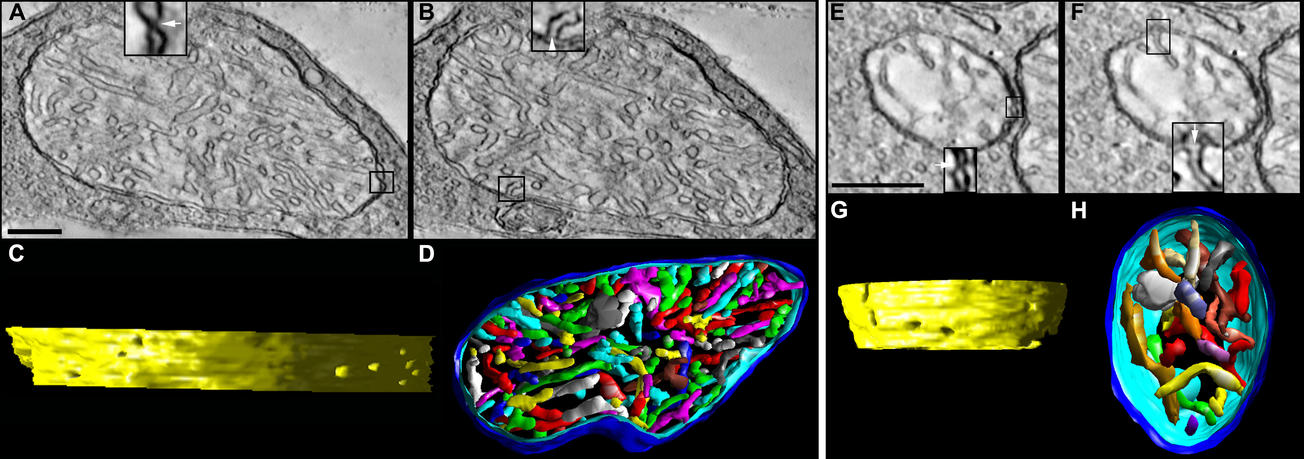
Figure 6. Bcl-xL does not fully protect spherule and pedicle mitochondria from postnatal lead exposure.
A–
D: Tomographic reconstructions of a Bcl-xL/Lead rod spherule mitochondrion from an adult mouse are presented. (
A and
B) The 2.2 nm slices through the middle of the volume show a large mitochondrion with fewer cristae compared to the control
and Bcl-xL and with contact sites and crista junctions (insets) with the same architecture as their control counterparts.
Scale bars=200 nm.
C: The side view of the inner membrane of the segmented volume is displayed with left lighting. There are nine numbered crista
junction openings in this view. The crista junction diameter is increased relative to the lead-exposed and control mitochondria;
see
Table 1.
D: The top view of the segmented volume shows the outer membrane and the entire complement of 97 cristae (various colors).
The cristae packing density is noticeably less than in the control or Bcl-xL mitochondria (compare to
Figure 3 and
Figure 4).
E–
H: Tomographic reconstructions of a Bcl-xL/Lead cone pedicle mitochondrion from an adult mouse are presented. (
E and
F) The 2.2 nm slices near the middle show a small mitochondrion. The contact sites and crista junctions (insets) have the same
architecture as the control, Bcl-xL, and lead. Scale bars=200 nm.
G: The side view of the inner membrane of the segmented volume shows eight crista junction openings in this view.
H: The top view of the segmented volume shows the outer membrane and the entire complement of 18 cristae (various colors).
The shape and size of the cristae are similar to that in the control and Bcl-xL mitochondria (compare to
Figure 3 and
Figure 4). However, the crista density is significantly less than in the lead-exposed mitochondrion (compare to
Figure 5).
 Figure 6 of
Perkins, Mol Vis 2012; 18:3029-3048.
Figure 6 of
Perkins, Mol Vis 2012; 18:3029-3048.