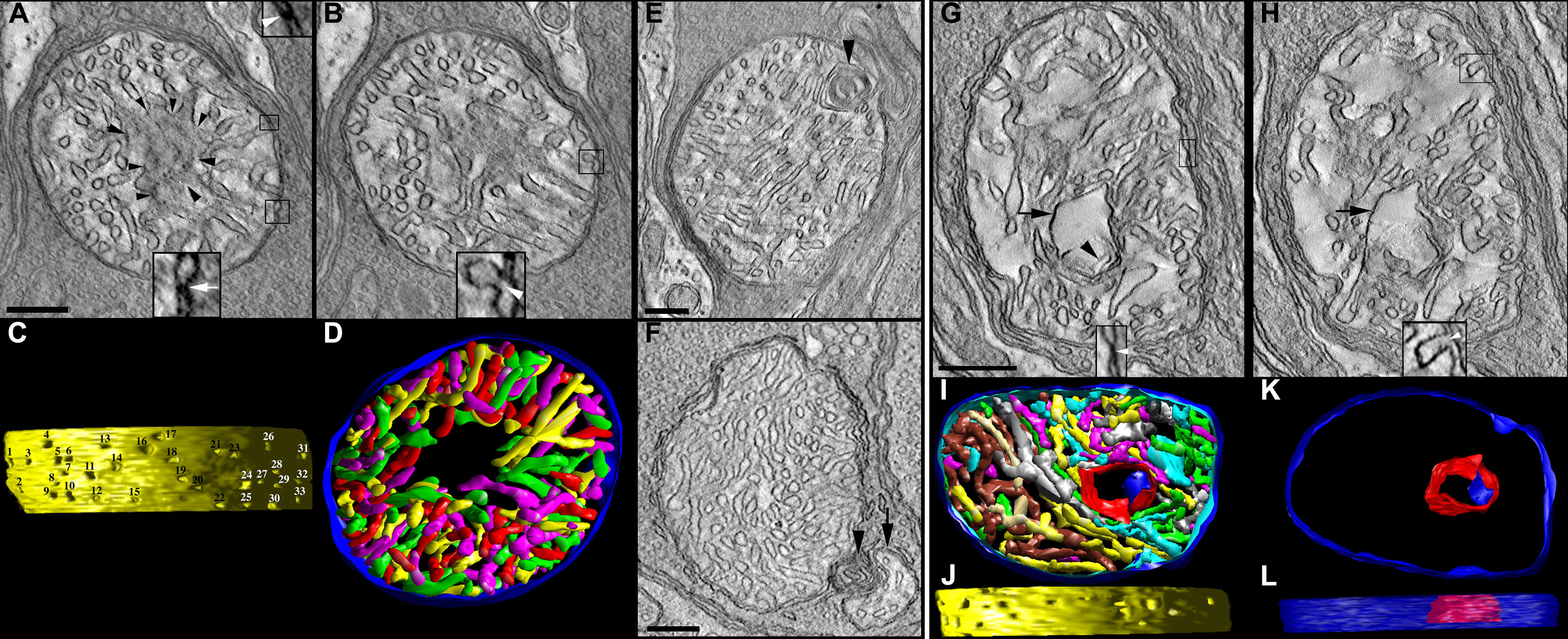
Figure 5. In the lead-exposed mice, the spherule and pedicle mitochondria have an altered cristae structure.
A–
D: Tomographic reconstructions of a rod spherule mitochondrion from a developmentally lead-exposed adult mouse are presented.
A: A 2.2 nm slice through the middle shows a medium-sized mitochondrion with cristae missing especially in the center of the
organelle (black arrowheads). The classic contact sites (arrow), bridge contact sites (arrowhead), and crista junctions (
B) have the same architecture as their control counterparts. Scale bars=200 nm.
B: Another slice through the volume shows the degraded cristae.
C: The side view of the inner membrane of the segmented volume is displayed with left lighting. There are 33 numbered crista
junction openings in this view.
D: The top view of the segmented volume shows the outer membrane and the entire complement of 98 cristae (various colors).
The region with missing cristae in the center of the mitochondrion extends throughout the reconstructed volume.
E: Another feature of lead-exposed mitochondria is a form of inner membrane degeneration known as “swirls” or “myelin-like”
(arrowhead).
F: A separate mitochondrion displayed the swirl (arrowhead) between the main mitochondrial body and a small mitochondrial fragment
(arrow) detached from the main body.
G–
L: Tomographic reconstructions of a cone pedicle mitochondrion from a developmentally lead-exposed adult mouse are presented.
(
G and
H) The 2.2 nm slices near the middle show a medium-sized mitochondrion containing a large, abnormal vesiculated crista (black
arrows) with a much smaller enclosed internal membrane (black arrowhead). As with the lead-exposed spherule mitochondrion,
the contact sites and crista junctions (insets) have the same architecture as the controls. Scale bars=200 nm.
I: The top view of the segmented volume shows the outer membrane and the entire complement of 69 cristae (various colors).
J: The side view of the inner membrane of the segmented volume shows 16 crista junction openings in this view. The crista junction
diameter is increased compared to the controls; see
Table 1.
K: The top view and (
L) side view of the vesiculated crista (red) and internal membrane (blue) show the extent of the volume (outer membrane in
translucent blue) occupied by this abnormal structure.
 Figure 5 of
Perkins, Mol Vis 2012; 18:3029-3048.
Figure 5 of
Perkins, Mol Vis 2012; 18:3029-3048.