![]() Figure 9 of
Mader, Mol Vis 2006;
12:915-930.
Figure 9 of
Mader, Mol Vis 2006;
12:915-930.
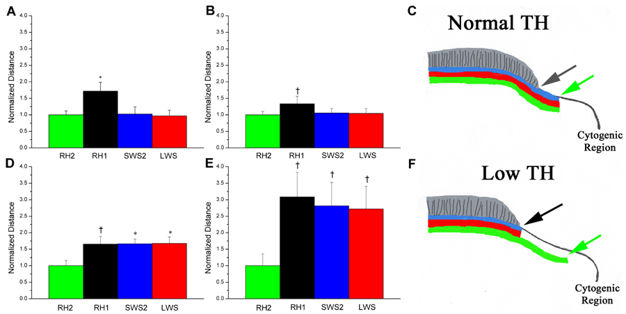
Figure 9. Opsin expression patterns at cytogenic regions of postmetamorphic flounder retina
Opsin expression was determined with in situ hybridization analysis of sequential sections as in Figure 7B, Figure 10, and Figure 11, and measurements between labeled photoreceptors and the cytogenic region were derived [32]. The measured distances for each opsin (mean±SD) were normalized with respect to the value for the RH2 opsin, and statistical analysis was performed with an independent Student's t-test (dagger indicated p<0.01; asterisk indicates p<0.05). A: At the CGZ of control retina RH1 expression is significantly spatially displaced from the RH2, LWS, and SWS2 cone opsins. There is no difference between the cone opsins. B: At the edge of regenerating lesion sites in control retina, the patterns of opsin expression are similar to that observed for the CGZ (Figure 8) [32]. C: A schematic of retinal opsin expression patterns in control postmetamorphic retina, relative to regions of cytogenesis (CGZ or lesion edge). The RH2, LWS, and SWS2 expression patterns at the ONL are represented by the green, red, and blue lines, respectively, and the location of rod outer segments is indicated in grey. The green arrow indicates the location of the RH2-expressing cone closest to the region of cytogenesis, which is spatially coincident with the corresponding LWS- and SWS2-expressing cones. D: At the CGZ of hypothyroidic retina there is a statistically significant, spatial displacement of LWS and SWS2 expression relative to RH2 expression, matching that of RH1 expression. E: At the edge of regenerating lesion sites in hypothyroidic retina the patterns of opsin expression are similar to those at the CGZ: a significant displacement of RH1, LWS, and SWS2 expression relative to RH2 expression. There is also a substantially greater displacement between the RH1 and RH2 expressing cells compared to control (Panel B). F: A schematic of retinal opsin expression patterns in hypothyroidic postmetamorphic retina, as in Panel C. The effects of induced hypothyroidism upon retinal growth include an absence of rod production (i.e., the greater displacement between RH1 and RH2 expression in Panel E compared to Panel B) and an absence of LWS and SWS2-expressing cones (i.e., a lack of correspondence between the ends of the green, red, and blue lines). With respect to the photoreceptor repertoire, retina grown or regenerated during hypothyroidic conditions phenotypically matches the retina of premetamorphic fish.