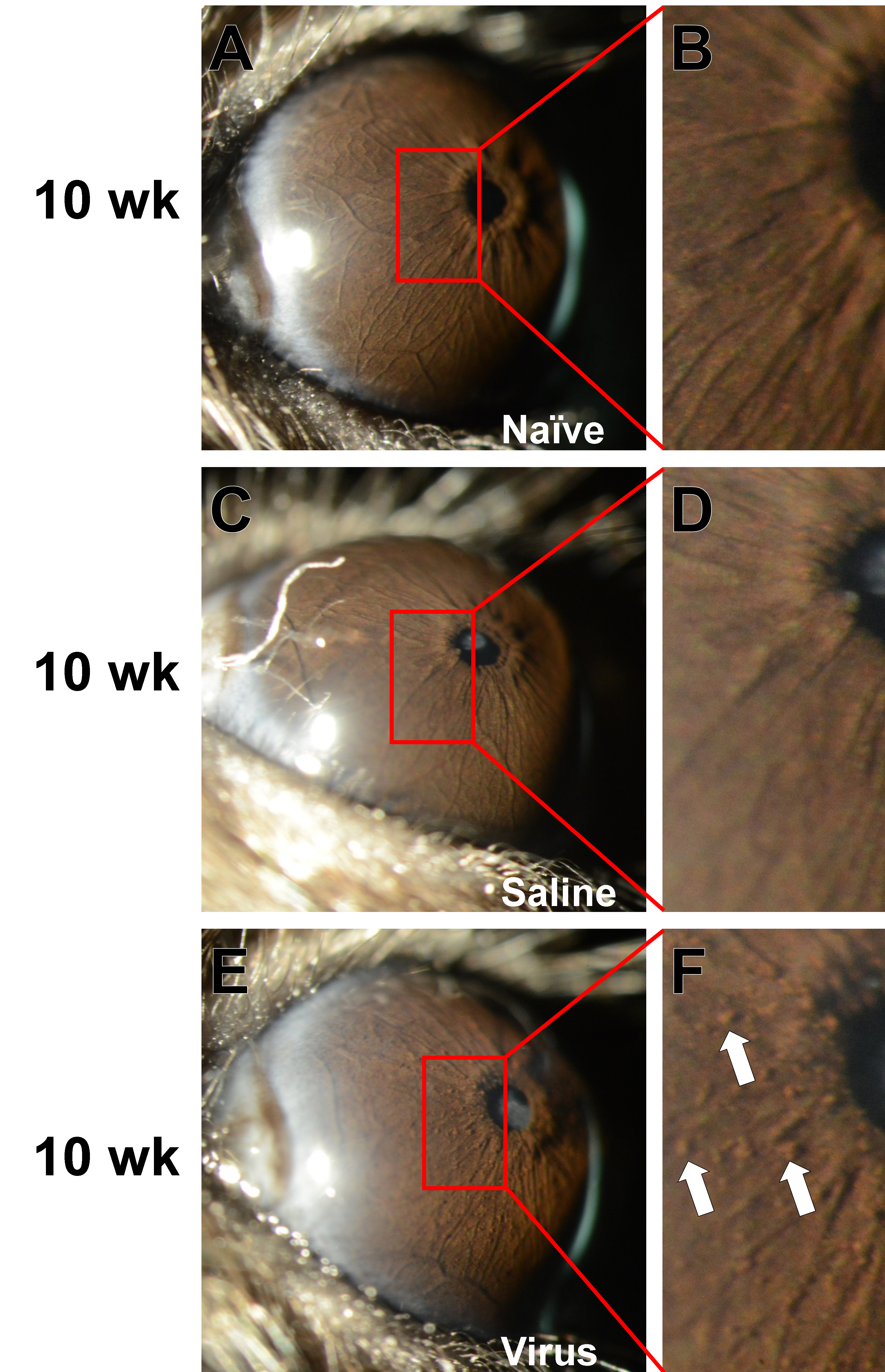
Figure 4. High-magnification slit-lamp images showing the unique presence of clump cells in type 5 adenovirus (Ad5)-injected eyes. Images
are from the same eyes shown at the 10-week time point in
Figure 1,
Figure 2 and
Figure 3 but photographed at a higher magnification and with the mouse held at a more severe angle with respect to the light source.
A digital enlargement of the same areas immediately to the left of each pupil is shown in the right-hand column.
A-B: The irides of mice in the Naïve group retained a normal morphology with no visible clump cells throughout the study. The
same eye is shown in
Figure 1G.
C-D: The irides of mice in the Saline group also maintained a normal morphology, continuing to lack visible clump cells. A lenticular
opacity is also visible. The same eye is shown in
Figure 2G.
E-F: Unique to treated eyes of the Virus group, intraocular injection of Ad5 led to a notable accumulation of clump cells (
white arrow, several additional cells are visible but unmarked) on the surface of the iris. Lenticular and corneal opacity is also apparent.
The same eye is shown in
Figure 3G. Images at 40X magnification were collected by an investigator who was masked to treatment status at the time the photographs
were taken.
 Figure 4 of
Meyer, Mol Vis 2021; 27:741-756.
Figure 4 of
Meyer, Mol Vis 2021; 27:741-756.