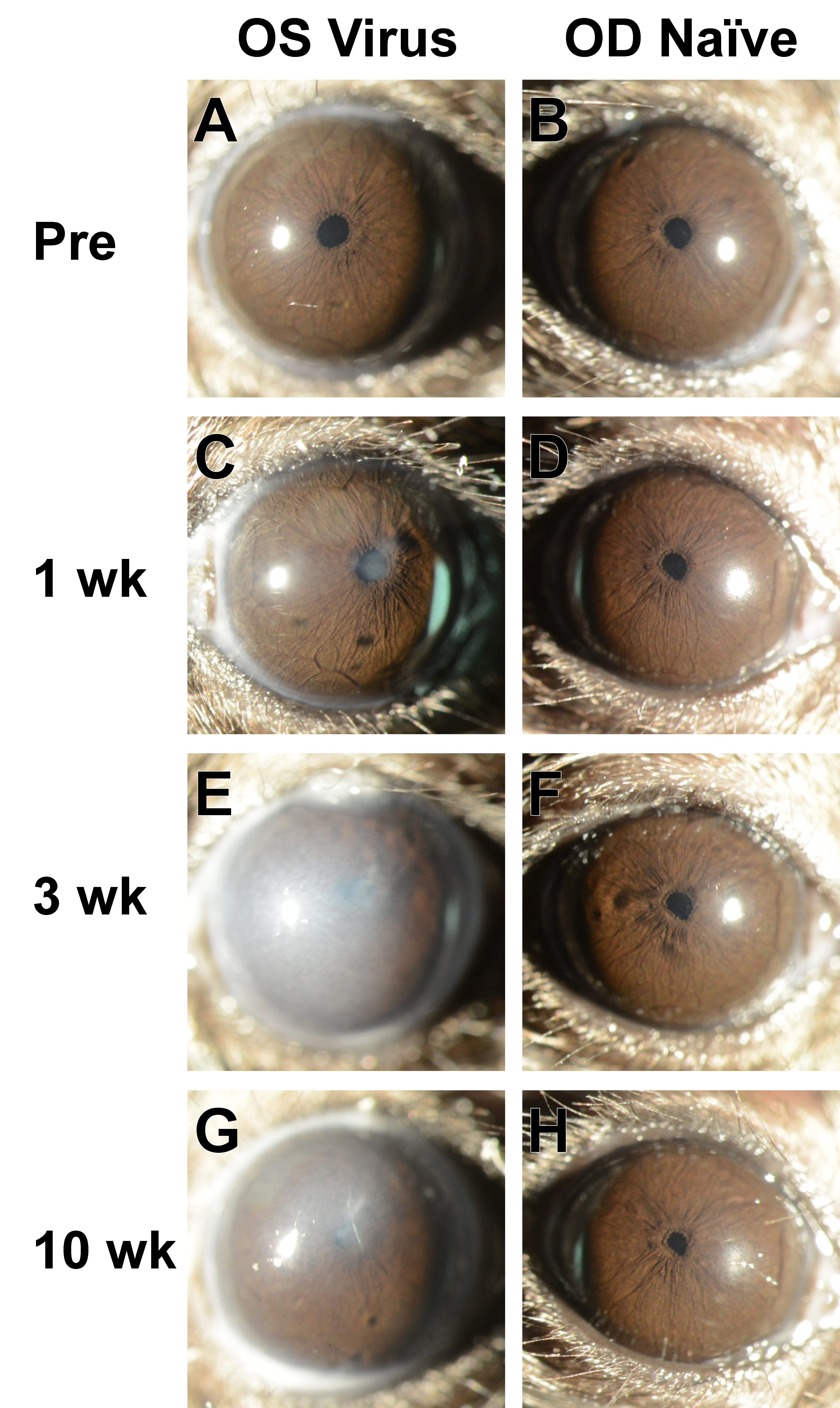
Figure 3. Slit-lamp images from a mouse in the Virus group. Images from the left and right eyes of a single mouse at progressive ages
showing consequences replication deficient species C type 5 adenovirus (Ad5) injection in the left eye and normal appearance
of the anterior chamber in the right eye.
A-B: Pretreatment images were collected in mice that were 12.5 weeks old. The left eye subsequently received an intraocular injection
of Ad5.
C-D: At the 1-week time point, the injected left eye has several areas showing mild corneal opacity and lenticular opacity; the
naïve right eye has a normal appearance.
E-F: At the 3-week time point, the corneal opacity of the injected left eye has become significantly more severe, blocking visualization
of the remainder of the anterior chamber; the uninjected right eye remains normal in appearance.
G-H: At the 10-week time point, the corneal opacity in the injected left eye shows lessened severity, and where areas of the
iris can be viewed, rough-appearing areas are present. The naïve right eye maintains a normal appearance. It should be noted
that the apparent cloudiness of the cornea also depends on the reflectivity of the light source; see
Figure 4E for a different view of the same eye and time point shown in panel
G. Images at 25X magnification were collected by an investigator who was masked to treatment status at the time the photographs
were taken.
 Figure 3 of
Meyer, Mol Vis 2021; 27:741-756.
Figure 3 of
Meyer, Mol Vis 2021; 27:741-756.