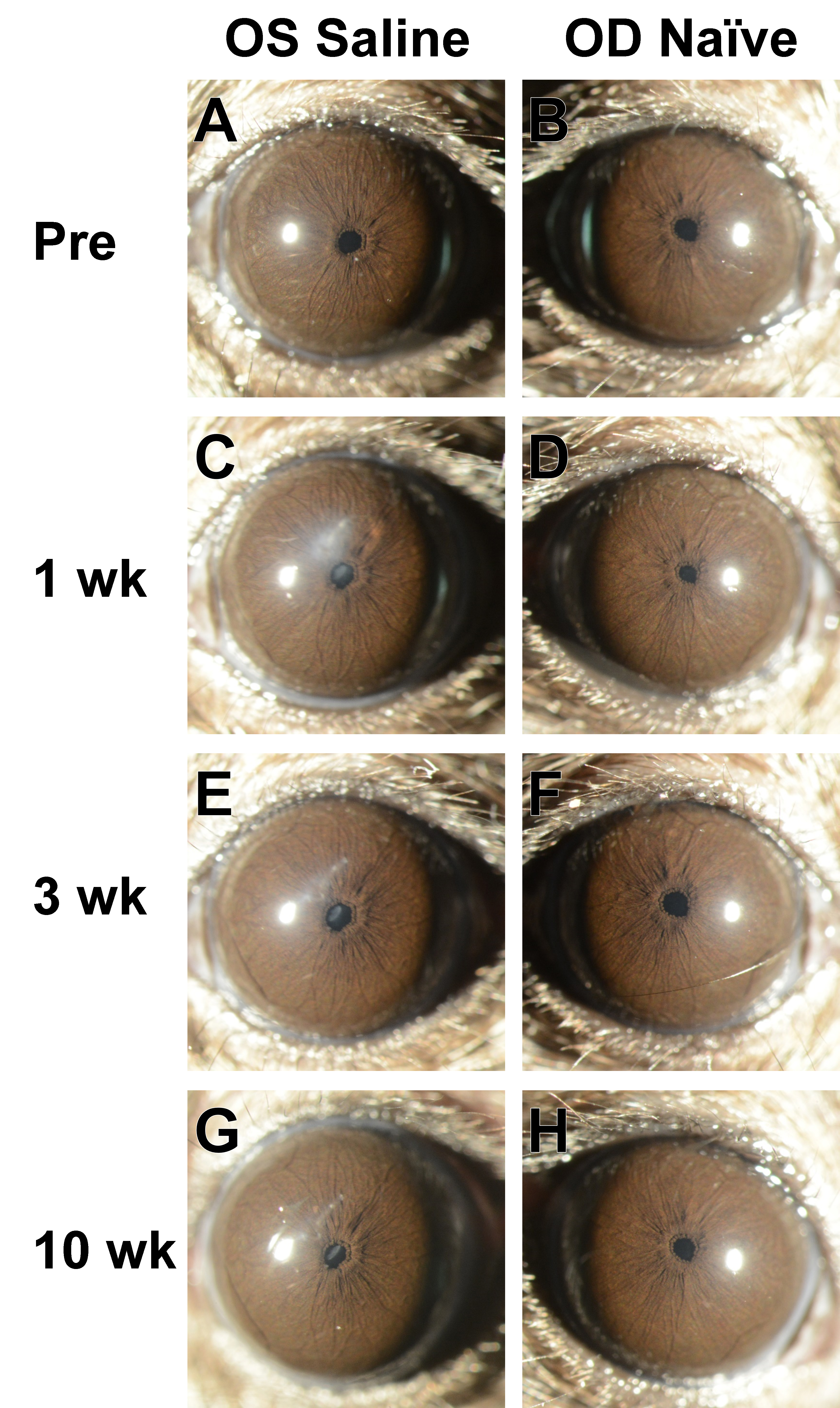
Figure 2. Slit-lamp images from a mouse in the Saline group. Images from the left and right eyes of a single mouse at progressive ages
show consequences of intraocular injection in the left eye and normal appearance of the anterior chamber in the right eye.
A-B: Pretreatment images were collected in mice that were 12.5 weeks old. The left eye was subsequently injected with balanced
salt solution (BSS).
C-D: At the 1-week time point, the injected left eye has a mild corneal opacity where the needle was inserted and slight lenticular
opacity; the naïve right eye has a normal appearance.
E-F: At the 3-week and
G-H: 10-week time points, the corneal opacity in the injected left eye becomes progressively less severe, and the lenticular
opacity appears to be unchanged; meanwhile, the naïve right eye maintains a normal appearance. See
Figure 4C for a different view of the same eye and time point shown in panel G. Images at 25X magnification were collected by an investigator
who was masked to treatment status at the time the photographs were taken.
 Figure 2 of
Meyer, Mol Vis 2021; 27:741-756.
Figure 2 of
Meyer, Mol Vis 2021; 27:741-756.