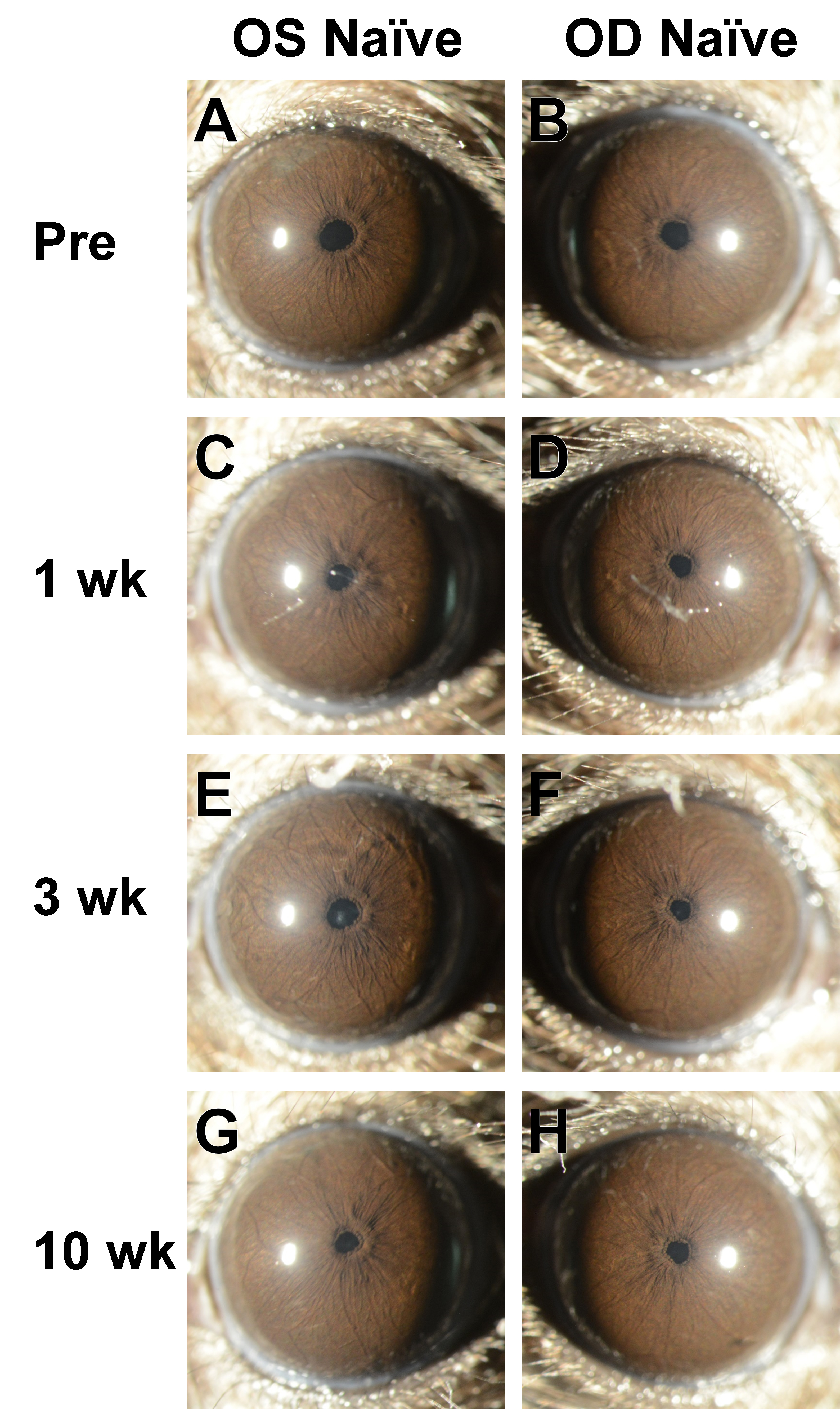
Figure 1. Slit-lamp images from a mouse in the Naïve group. Images from the left and right eyes of a single mouse at progressive ages
showing normal appearance of the anterior chamber.
A-B: Pretreatment images were collected in mice that were 12.5 weeks old. The cornea is clear, and the iris vessels are the main
notable feature of the iris. With subsequent aging in these unmanipulated mice, the same healthy appearance is maintained
at
C-D: 1 week,
E-F: 3 weeks, and
G-H: 10 weeks following initiation of the experiment. See
Figure 4A for a different view of the same eye and time point shown in panel G. Images at 25X magnification were collected by an investigator
who was masked to treatment status at the time they were photographed.
 Figure 1 of
Meyer, Mol Vis 2021; 27:741-756.
Figure 1 of
Meyer, Mol Vis 2021; 27:741-756.