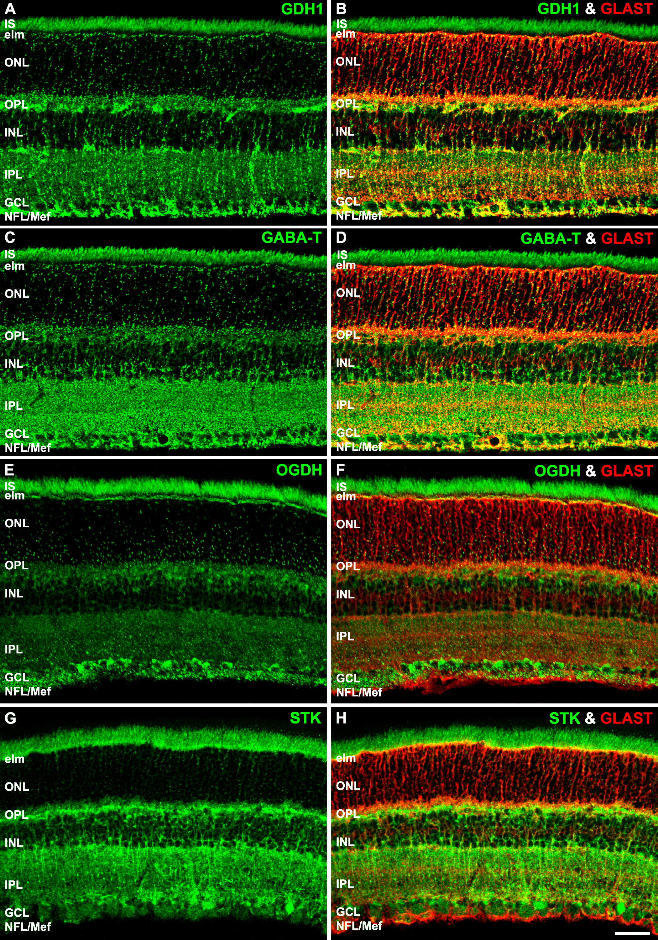
Figure 10. Confocal images reveal a unique metabolic signature of Müller cell mitochondria.
A: Retinas immunolabeled for glutamate dehydrogenase 1 (GDH1) reveal intense expression in rod and cone ISs and synaptic terminals,
and a strong to intense in the ELM, IPL, GCL, and MGC end-feet. The expression in the ONL is punctate and strong to intense.
B: Strong to intense colocalization of GDH1-IR and glial high-affinity glutamate-aspartate transporter (GLAST-IR) is seen throughout
the MGCs.
C: Retinas immunolabeled for GABA-transaminase (GABA-T) reveal an intense expression in rod and cone ISs and synaptic terminals,
and a strong to intense in the ELM, IPL, GCL, and MGC end-feet. The expression in the ONL is punctate and strong to intense.
D: Strong to intense colocalization of GABA-T-IR and GLAST-IR is seen throughout the MGCs.
E: Retinas immunolabeled for the lipoamide subunit of the 2-oxoglutarate (α-ketoglutarate) dehydrogenase complex (OGDH) reveal
intense expression in the rod and cone IS and synaptic terminals and a strong to intense in the ELM, IPL, GCL, and MGC end-feet.
The expression in the ONL is punctate and strong to intense.
F: OGDH-IR and GLAST colocalization is strong in the ELM and end-feet, and moderate distal and proximal processes, OPL, and
somas.
G: Retinas immunolabeled for succinate thiokinase (STK) reveal intense expression in the rod and cone ISs and synaptic terminals
and strong to intense expression in the ELM, IPL, GCL, and MGC end-feet. The expression in the ONL is moderate and more like
that of COX IV-IR (
Figure 4,
Figure 5, and
Figure 7).
H: STKH-IR and GLAST colocalization is strong in the ELM, proximal processes, and end-feet and moderate in the distal processes,
OPL, and somas. COX IV-IR = cytochrome oxidase IV immunoreactivity, ELM = external limiting membrane, GCL = ganglion cell
layer, INL = inner nuclear layer, IPL = inner plexiform layer, ISs = inner segments, MGC = Müller glial cell, NFL = nerve
fiber layer, ONL = outer nuclear layer. A–H, scale bars = 40 µm.
 Figure 10 of
Rueda, Mol Vis 2016; 22:847-885.
Figure 10 of
Rueda, Mol Vis 2016; 22:847-885.