Figure 7. Confocal images show that pan-LDH and LDH isozyme 5 (LDH-5: liver and muscle type) are expressed in almost all retinal cells
and synapses. A: Retina immunolabeled for pan-LDH. B: Retinas double-labeled for pan-LDH and VGLUT1 show colocalization in the OPL and the IPL (aquamarine pixels). C: Retinas double-labeled for pan-LDH and glutamine synthetase (GS) show weak colocalization in the MGC somas and ELM. D: Retina immunolabeled for LDH-5. E: Retinas double-labeled for LDH-5 and VGLUT1 show colocalization in the OPL and the IPL (aquamarine pixels). F: Retinas double-labeled for LDH-5 and COX IV show collocation in the ISs, OPL, and IPL. Inset: Single and double-labeled
cone ISs from a high-magnification image revealing colocalization in the ISs. G: High-magnification image of a retina double-labeled for LDH-5 and calbindin shows no colocalization. H: High-magnification image of a retina double-labeled for LDH-5 and CHX10 reveals differential colocalization. I: High-magnification image of a retina double-labeled for LDH-5 and PKCα shows colocalization in the somas but not in the
dendrites. COX IV = cytochrome c oxidase subunit IV, ELM = external limiting membrane, IPL = inner plexiform layer, IPL-a
= IPL sublamina-a, IPL-b = IPL sublamina-b, ISs = inner segments, LDH = lactate dehydrogenase, MGC = Müller glial cell, ONL
= outer nuclear layer, OPL = outer plexiform layer, OSs = outer segments, PKCα-IR = protein kinase C α immunoreactivity, VGLUT1
= vesicular glutamate transporter 1. A–F, scale bar = 40 µm. G–I, scale bar = 20 µm.
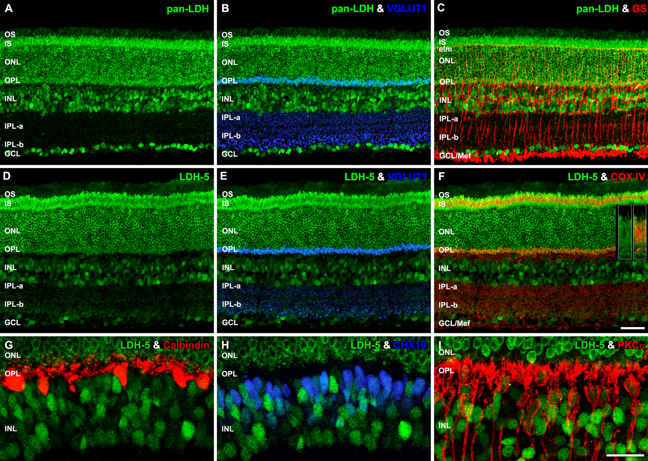
 Figure 7 of
Rueda, Mol Vis 2016; 22:847-885.
Figure 7 of
Rueda, Mol Vis 2016; 22:847-885.