Figure 5. Confocal images reveal that PFK-L1 is expressed in all retinal cells and synapses. A: Retina immunolabeled for phosphofructokinase isoform L1 (PFK-L1). B: Retinas double-labeled for PFK-L1 and VGLUT1 show colocalization in the OPL and the IPL (aquamarine pixels). C: Retinas double-labeled for PFK-L1 and COX IV show intense colocalization in the IS and the OPL regions. High-magnification
insets of highlighted cone shows single- and double-labeled colocalization in the OS, IS, and ONL of the perinuclear mitochondrion.
COX IV = cytochrome c oxidase subunit IV, ISs = inner segments, IPL = inner plexiform layer, IPL-a = IPL sublamina-a, IPL-b
= sublamina-b, ONL = outer nuclear layer, OPL = outer plexiform layer, OSs = outer segments, VGLUT1 = vesicular glutamate
transporter 1. A and B, scale bar = 40 µm. C, scale bar = 20 µm.
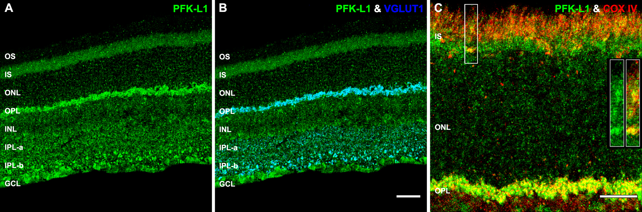
 Figure 5 of
Rueda, Mol Vis 2016; 22:847-885.
Figure 5 of
Rueda, Mol Vis 2016; 22:847-885.