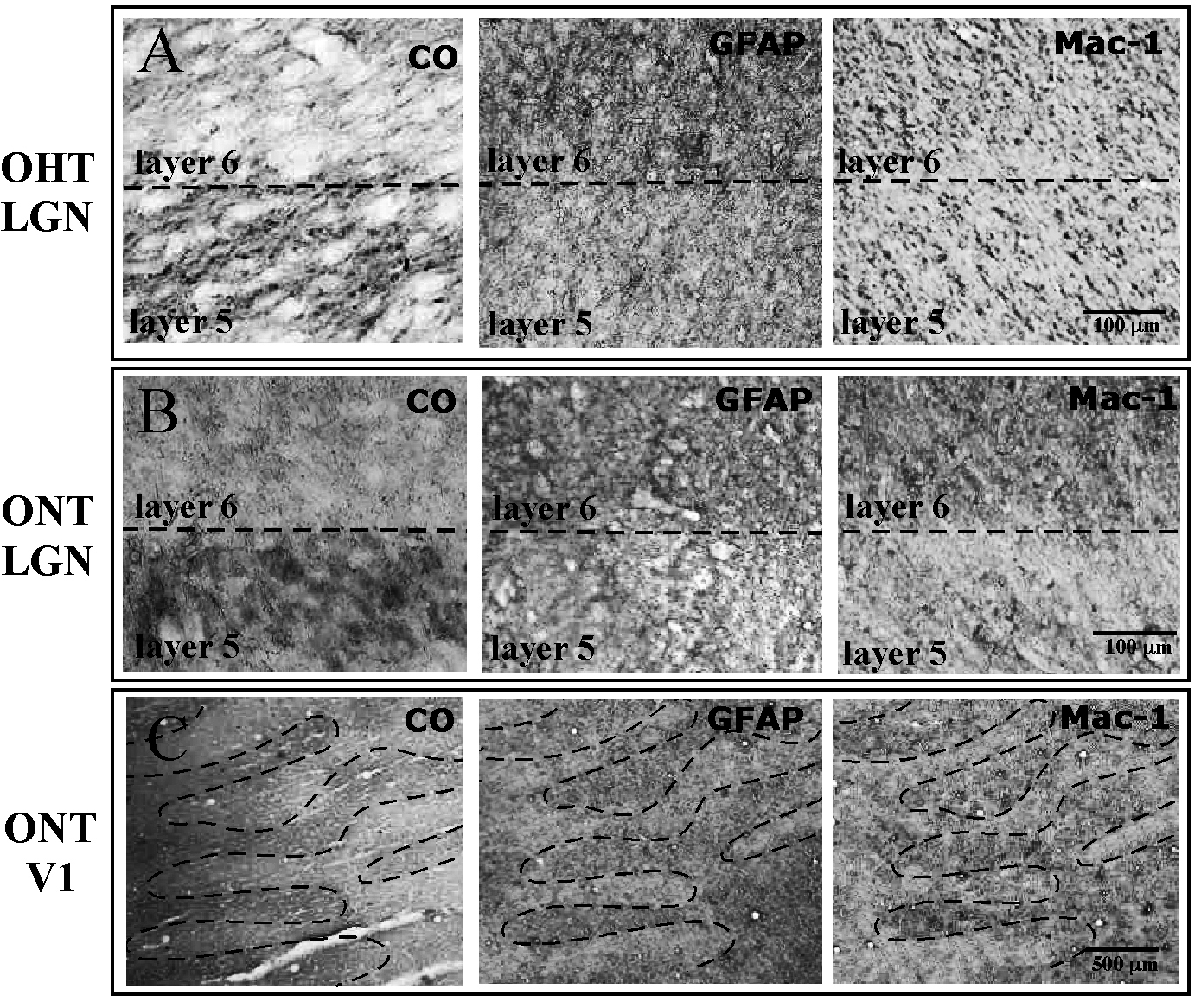
Figure 6. CO histochemistry, GFAP and MAC-1 immunohistochemistry in unilateral ONT and OHT brains. After 14 days, the CO staining demonstrated
lighter staining in the contralateral (treated) eye bands (Layer 1, 4, 6) in the LGN of both the OHT (534) and the ONT (32676)
brains, consistent with the findings from animals with vision loss of longer duration (
Figures 1–
4). The GFAP immunoreactivity was stronger in the regions of LGN and V1 associated with the treated eye in both OHT (
A) and ONT (
B,
C) brains. The MAC-1 immunoreactivity was stronger in the regions associated with the treated eye in ONT (
B,
C) brains only. MAC-1 immunoreactivity did not show differential expression between treated and normal ocular dominance bands,
consistent with findings from long-term OHT (
Figure 3 and
Figure 4).
 Figure 6 of
Lam, Mol Vis 2009; 15:2217-2229.
Figure 6 of
Lam, Mol Vis 2009; 15:2217-2229.