Figure 1. Photomicrographs of serial
coronal sections of LGN from an ONT brain. A: Section stained
for CO histochemistry demonstrates reduced CO activity in contralateral
(Layers 1,4,6) eye layers. B: Serial section stained for GFAP
immunohistochemistry demonstrates denser reaction product in the
contralateral eye layers. C: Serial sections stained for MAC-1
immunohistochemistry demonstrates a denser staining pattern in the
contralateral eye layers. Arrows point to high power photographs (100X)
of treated versus normal LGN layers stained for CO (D, E),
GFAP (F, G), or MAC-1 (H, I). Note
astrocytic profiles and processes in F and microglial profiles
in H.
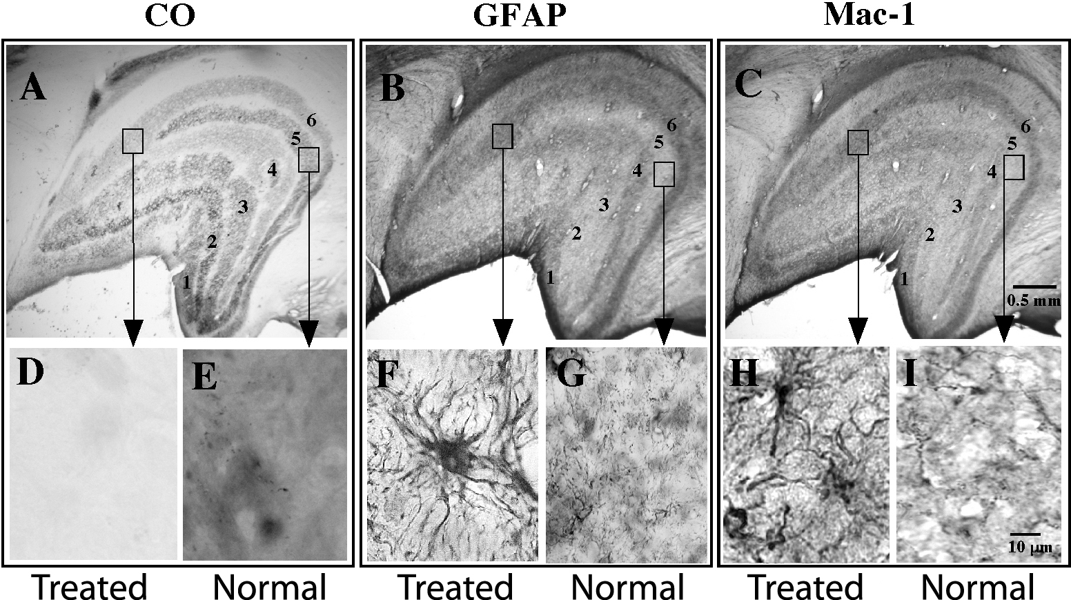
 Figure 1 of Lam, Mol Vis 2009; 15:2217-2229.