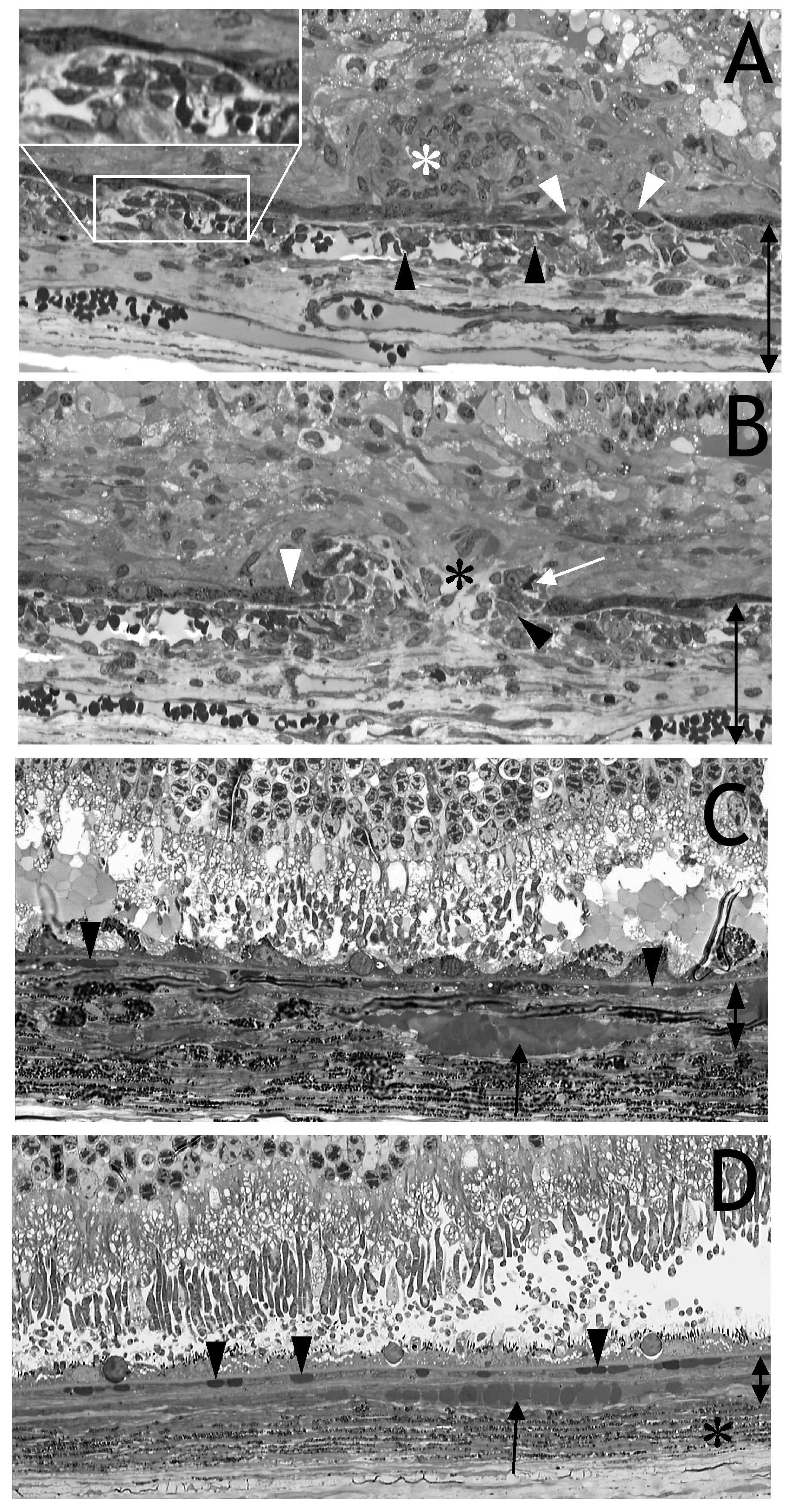
Figure 4. Semithin sections of the eye
shown in
Figure
2J,K.
A: The endothelial cell layer of the
choriocapillaris was irregular, and endothelial cells protruded into
the vessel lumen (black arrowheads). Evidence for this is presented by
electron microscopy (see
Figure 5A-C). The retinal pigment
epithelium (RPE) cell layer was disrupted (white arrowheads), and
endothelial cells migrated and proliferated into the subretinal space
or between Bruch’s membrane and RPE (inset). Evidence that these cells
were endothelial cells is presented by immunohistochemistry (see
Figure 6A).
These cellular proliferations were either solid (white asterisk in
A)
or loosely packed with interstitial spaces (black asterisk in
B).
The photoreceptors have already degenerated, and retinal scar was
closely connected to the RPE and proliferating cells. This was probably
why fluorescein leakage was restricted to the spotted roundish areas
visible in
Figure
2. An immature capillary containing an erythrocyte was
located distally to the RPE (
B, white arrow). The melanocytes of
the choroids were located below the deeper choroidal vessels and are
not shown.
C and
D: After injection of HC Ad. EGFP or
PBS, the RPE, choriocapillaris (arrowheads) and deeper choroidal
vessels (arrow) appeared to be normal. The pigmented layer, consisting
predominately of melanocytes, is marked by a black asterisk. The double
arrows in
A-C indicate growth of extracellular matrix and
vessel layers of the choroid after VEGF expression (
A, B)
compared to (
C).