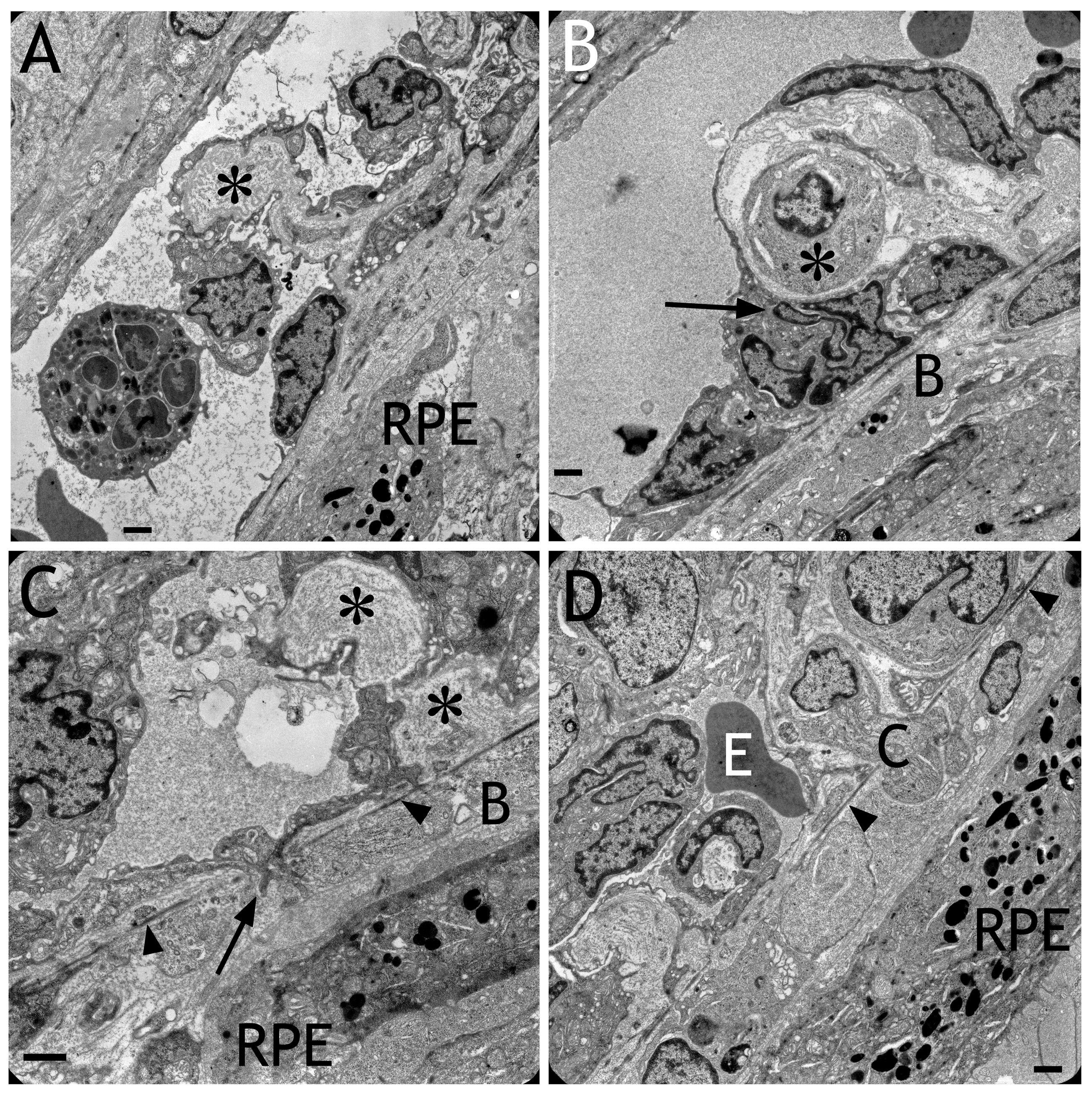
Figure 5. Electron micrographs obtained
after transduction with HC Ad.VEGF-A.
A: An invagination of the
the endothelium into the lumen of the choriocapillaris containing
extracellular matrix (asterisk) is shown. This caused the patchy
appearance of the choriocapillaris lumen presented in
Figure 4.
B:
A cell (asterisk) was located between the endothelium and Bruch’s
membrane. Note the extremely frayed or fragmented nucleus of an
endothelial cell (arrow).
C: An endothelial cell was spreading
into Bruch’s membrane (B) toward the RPE (arrow). The elastic layer of
Bruch’s membrane is labeled by arrowheads. Sites indicating remodeling
of the extracellular matrix surrounding the choriocapillaris are
labeled by asterisks.
D: A cell (C) was migrating into Bruch’s
membrane toward the RPE. The elastic layer of Bruch’s membrane is
labeled by arrowheads. Within the pathological capillary, an
erythrocyte (E) was still present. Scale bars in each image: 1 μm.