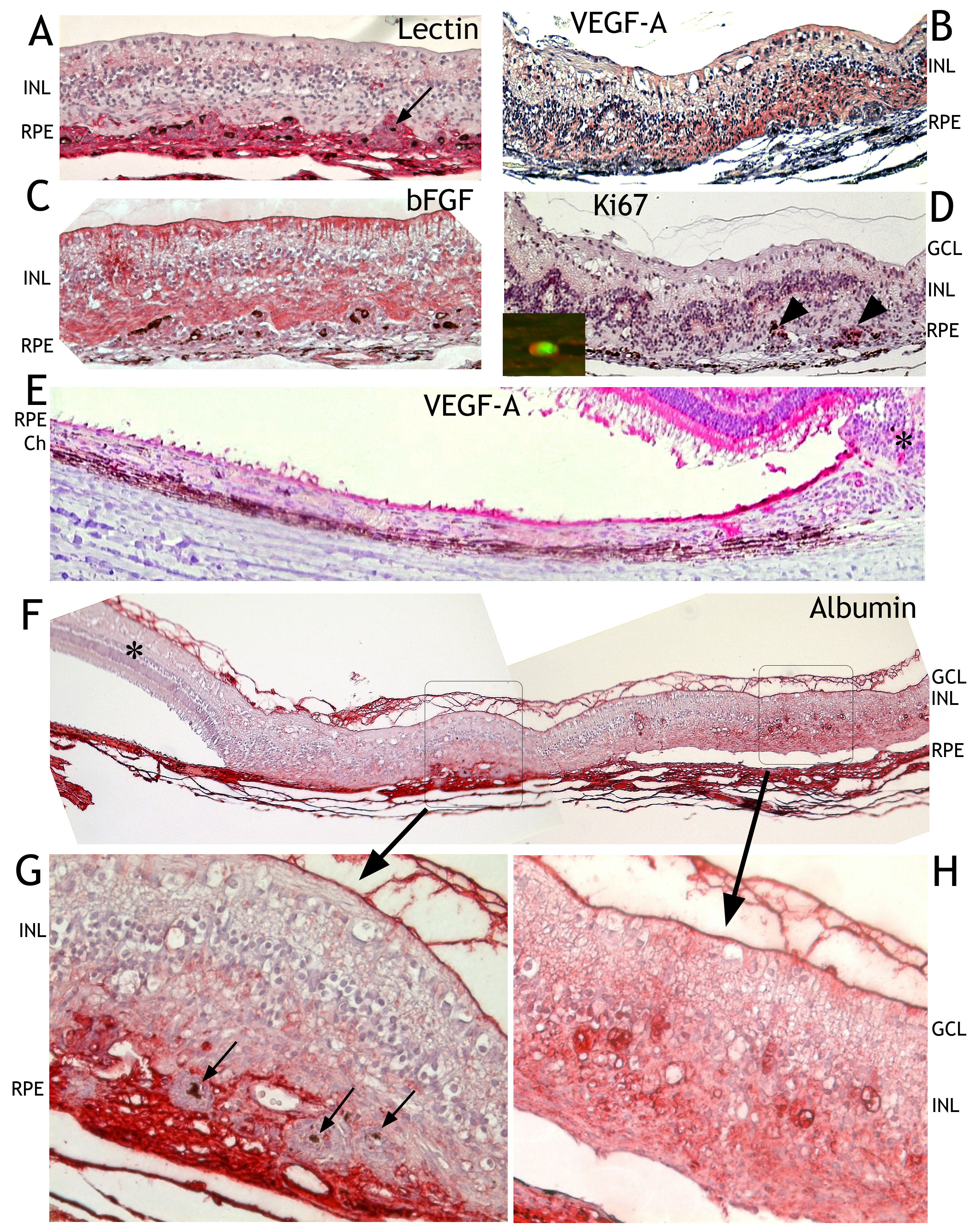
Figure 6. Immunohistochemical findings
after transduction with HC Ad.VEGF-A with hematoxylin and eosin
counterstain.
A: Staining with tomato lectin showed that most
cells penetrating the disrupted retinal pigment epithelium (RPE) cell
layer were of endothelial origin (red). Inner and outer retinal nuclear
layers were mixed. Single RPE cells (black arrow) were surrounded by
proliferating endothelial cells.
B: VEGF-A was highly expressed
in the retinal scar (white asterisk) and in the RPE close to the
retinal scar (black asterisk) but this expression rapidly decreased the
farther it was from the retinal scar (
E). The expression of bFGF
was not as strong in cells of choroidal origin than in retinal cells (
C).
D: Proliferating cells at the retinal choroidal interface were
immunoreactive for Ki67 (black arrowheads). The inset in (
D)
demonstrates the endothelial nature of dividing cells by double
labeling for Ki67 (red) and tomato lectin (green).
F: Albumin
(red) was present in the choroid and the fiber matrix at the
vitreoretinal interface. It was absent in the unaffected retina (black
asterisk). Two selected areas are shown enlarged (
G, H).
Choroidal retinal scarring and leakage of albumin are visible in one of
the enlarged areas (
G). Albumin was not localized within
proliferating cells surrounding single RPE cells (black arrows). This
may be why ICG did not enter such sites (see
Figure 2I,M).
H: The second
enlarged area revealed albumin leakage within the retina that, as shown
in this panel, was not fused with RPE or choroid (Ch). Abbreviations:
INL, inner nuclear layer; GCL, ganglion cell layer.