![]() Figure 7 of
Beattie, Mol Vis 2005;
11:825-832.
Figure 7 of
Beattie, Mol Vis 2005;
11:825-832.
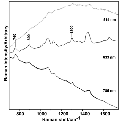
Figure 7. Raman spectra of enriched porcine rhodopsin
Raman spectra of enriched porcine rhodopsin preparation (purified by HPLC) acquired using 785, 633, and 514 nm excitation. Some bands which are unusually intense for a protein are labeled; these unique bands do not occur in the Raman spectra of the POS recorded at the same three excitation wavelengths (Figure 1, Figure 3, Figure 4). The baseline variation is non-Raman in origin.