![]() Figure 4 of
Jablonski, Mol Vis 2001;
7:27-35.
Figure 4 of
Jablonski, Mol Vis 2001;
7:27-35.
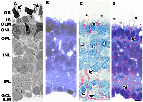
Figure 4. Lactose supported glutamine synthetase expression: morphology and immunolocalization patterns.
A: Lactose permitted organization of the photoreceptor outer segments in the absence of the RPE (arrows) and cell loss was minimized. B: GFAP expression was suppressed in contrast to the upregulation noted in RPE-deprived retinas (compare to Figure 2B) C: CRALBP immunolabeling was heavy, following Müller cell processes (arrow) and ending at the outer and inner limiting membranes (black arrowheads). Occasional INL nuclei were outlined (white arrows). Some immunopositive label was present in the interphotoreceptor space, quite distal from the outer limiting membrane (asterisks). D: Glutamine synthetase expression was maintained in the presence of lactose. Immunopositive labeling was present across the entire retina. Heavier immunolabeling was found over the plexiform layers (arrows), similar to control conditions. In addition, glutamine synthetase immunoreactiviity was present in an area around the inner segment/outer segment junction (asterisks), similar to the localization of CRALBP shown in Figure 3C. RPE = retinal pigment epithelium, OS = outer segment, IS = inner segment, OLM = outer limiting membrane, ONL = outer nuclear layer, OPL = outer plexiform layer, INL = inner nuclear layer, IPL = inner plexiform layer, GCL = ganglion cell layer, ILM = inner limiting membrane. Bar = 10 mm.
