![]() Figure 2 of
Jablonski, Mol Vis 2001;
7:27-35.
Figure 2 of
Jablonski, Mol Vis 2001;
7:27-35.
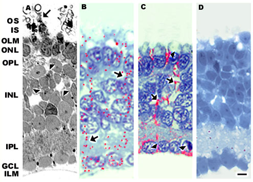
Figure 2. Morphology and immunolocalization patterns of Müller cell-specific proteins
Morphology and immunolocalization patterns of Müller cell-specific proteins in retinas induced to degenerate by removal of the RPE. A: In the absence of the RPE, photoreceptor outer segments were elaborated as whorl-like structures (arrows). Cell loss was apparent in both the ONL and INL (arrowheads). B: GFAP expression was present across the entire retina (arrows) from outer limiting membrane to inner limiting membrane. C: CRALBP labeled the entire Müller cell (arrows) and ended abruptly at the limiting membranes (arrowheads). Müller cell nuclei were not outlined and no immunolabeling was present beyond the outer limiting membrane, as seen in control eyes. D: Glutamine synthetase expression was completely suppressed in these retinas. RPE = retinal pigment epithelium, OS = outer segment, IS = inner segment, OLM = outer limiting membrane, ONL = outer nuclear layer, OPL = outer plexiform layer, INL = inner nuclear layer, IPL = inner plexiform layer, GCL=ganglion cell layer, ILM = inner limiting membrane. Bar = 10 mm.