![]() Figure 3 of
Riley, Mol Vis 2001;
7:297-304.
Figure 3 of
Riley, Mol Vis 2001;
7:297-304.
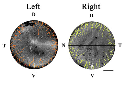
Figure 3. Quadrant definitions of left and right corneas
E16 Japanese quail corneas from left and right sides were immunostained for nerves as in Figure 1, with images captured via confocal microscopy (from endothelial side), digitized, and imported into Adobe Photoshop 5.5. Each cornea was divided into four equal quadrants, using the ventral pole marker (choroid fissure) as a reference point. Each nerve entrance point in each quadrant was counted, allowing quantitative comparison of innervation of each quadrant of left and right corneas (Table 1 and Table 2). Abbreviations: D, dorsal; N, nasal; T, temporal; V, ventral. Bar represents 500 mm.