![]() Figure 1 of
Riley, Mol Vis 2001;
7:297-304.
Figure 1 of
Riley, Mol Vis 2001;
7:297-304.
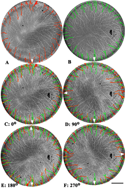
Figure 1. Overlays and rotations of corneal nerve images
E16 Japanese quail cornea whole mounts were immunostained with monoclonal antibody QN for quail nerves, with images captured via confocal microscopy (from the endothelial side), digitized and imported into Adobe Photoshop 5.5 for nerve visualization. Nerve entrance points were made distinct from background by choosing the contrasting pseudo-colors of the nerve images (e.g., red or green) and subsequently graying the image of the rest of the corneal image (A and B). Superposition test: Corneal images from the same side were then overlaid, with the ventral pole marker (choroid fissure; arrow) of each cornea placed in register (C). Where nerves from different corneas are in exactly the same positions, a third pseudo-color (yellow) is formed. Rotation test: Subsequent 90° rotations of one corneal image with respect to the other were made to test for diminishing positional specificity, assuming the original nerve entry points were at specific locations (D, E, and F). However, the degree of entry position synchrony, as revealed by the appearance of the third pseudo-color (yellow), does not significantly decrease, suggesting that corneal innervation does not occur at specific locations, but rather occurs at rather uniform spaced sites around the entire circumference of the cornea. Bar represents 500 mm.