![]() Figure 4 of
Cai, Mol Vis 2000;
6:132-143.
Figure 4 of
Cai, Mol Vis 2000;
6:132-143.
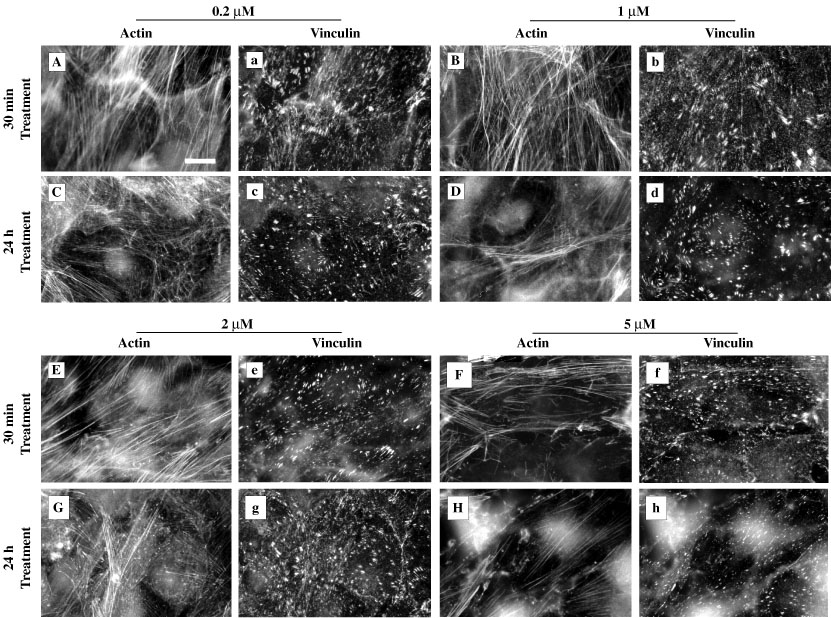
Figure 4. Effect of LAT-A on the distribution of actin and vinculin in cultured HTM cells
Effect of LAT-A on the distribution of actin and vinculin in cultured HTM cells treated with 0.2, 1, 2, or 5 mM LAT-A for 30 min and 24 h, and double-labeled for actin (panels A-H) and vinculin (panels a-h). See Figure 3A and Figure 3B for normal actin and vinculin distribution and Figure 6 for actin and vinculin distribution after LAT-A treatment for 2 h. LAT-A resulted in progressive deterioration of actin filaments, and time- and dose-dependent disorganization of vinculin-containing focal contacts. The latter appeared more resistant to LAT-A than the b-catenin-containing intercellular adhesions, even at higher doses and longer treatment duration (panels g and h; compare with Figure 5). Bar = 20 mm.