![]() Figure 3 of
Cai, Mol Vis 2000;
6:132-143.
Figure 3 of
Cai, Mol Vis 2000;
6:132-143.
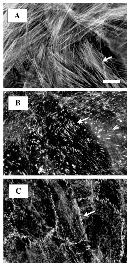
Figure 3. Distribution and organization of actin filaments and actin-related proteins in normal HTM cells
Fluorescent staining for actin revealed numerous stress fibers oriented primarily parallel to the long axis of the cells (A) and terminating at vinculin-containing focal adhesions and adherens junctions (A, B). Panels A and B are the same cells. Intercellular junctions were delineated by b-catenin staining appearing as continuous or partially segmented lines at cell-cell borders (C). The arrows point to actin stress fibers (A), vinculin-containing focal adhesions (B), and b-catenin containing cell-cell junctions (C). Bar = 30 mm.