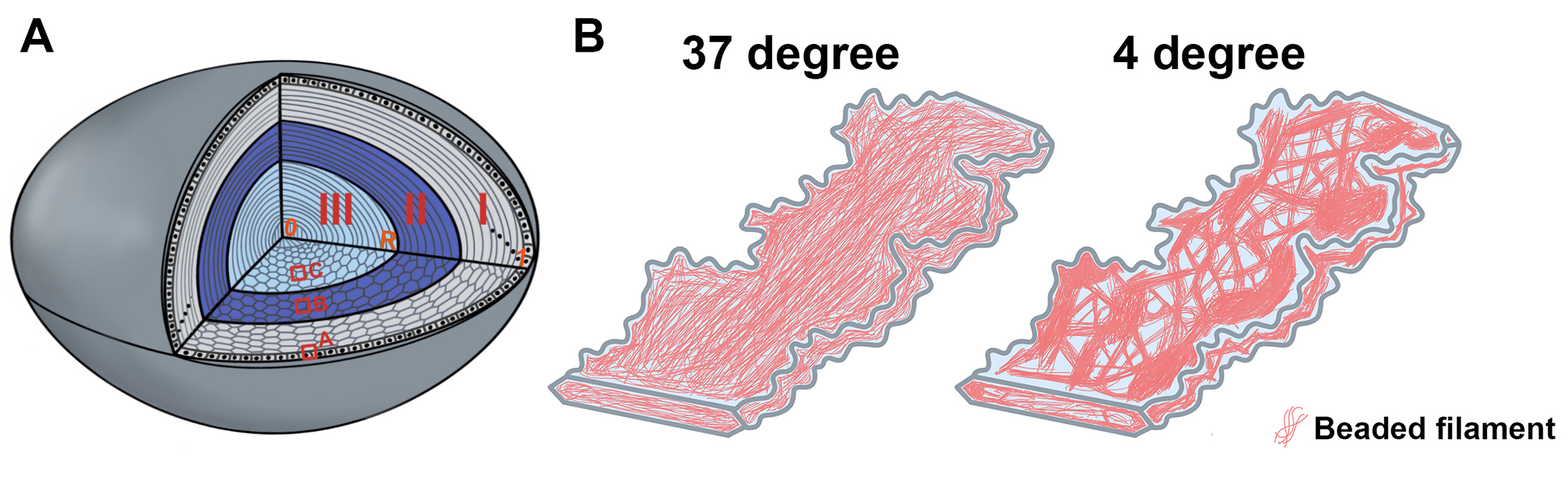
Figure 5. Illustration of lens structures and changes of intermediate filaments in the formation of cold cataract.
A: A schematic lens model shows three zones (I, II, and III) according to the 25 °C induced cold cataract in zone III and the
4 °C degree induced cold cataract in zone II and zone III in P14 B6 wild-type lenses. The red boxes (
A,
B, and
C) indicate the locations of the lens images in
Figure 2,
Figure 3, and
Figure 4. The radius (R) of the P14 lens from the lens capsule (1) to the lens center (0) is about 875 μm. Zone I fibers (gray) cover
0–245 μm in depth, zone II (dark blue), 245–420 μm in depth, and zone III (light blue), 420–875 μm in depth from the lens
surface.
B: An interior hexagonal fiber cell model shows the change in the CP49 and filensin beaded intermediate filaments from uniform
distribution at 37 °C to intermittent accumulation at 4 °C.
 Figure 5 of
Li, Mol Vis 2020; 26:603-612.
Figure 5 of
Li, Mol Vis 2020; 26:603-612.