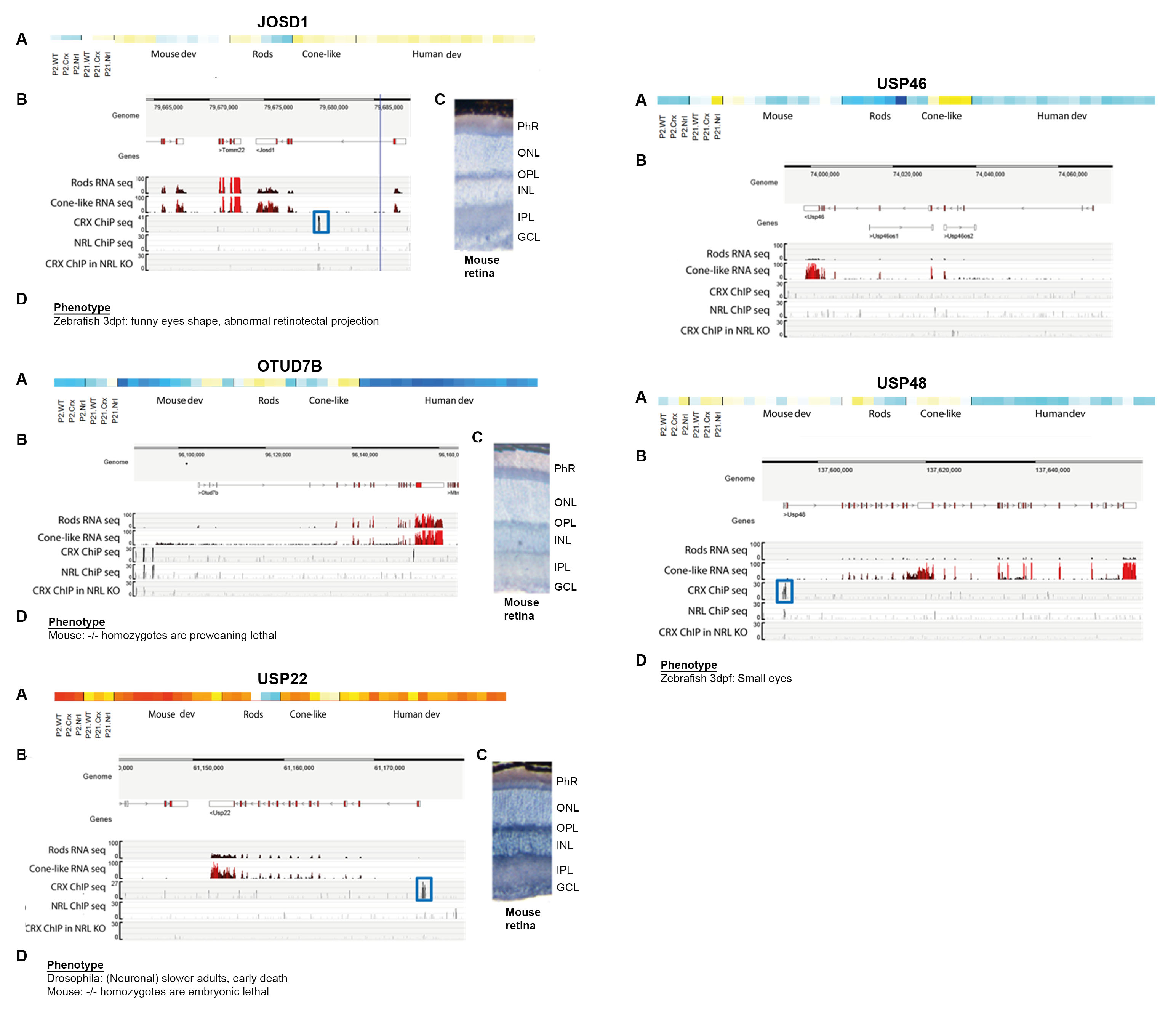
Figure 3. Diagram showing the criteria considered for the selection of five relevant DUB genes in the retina. For each of the selected
genes,
Josd1, Otud7b, Usp22, Usp46, and
Usp48, the composite images show the following.
A: Transcriptome analysis of each selected gene, extracted from
Figure 2.
B: Track view of CRX- and NRL-chromatin immunoprecipitation sequencing (ChIP-seq) density profiles (after CRX and NRL immunoprecipitation,
respectively) using postnatal day 28 (P28) wild-ty[e (WT) mouse retinas, visualized using the UCSC genome browser (
https://neicommons.nei.nih.gov/).
C: Pattern of expression with in situ hybridization in WT mouse retinas (reported in [
30]). PhR, photoreceptor cell layer; ONL, outer nuclear layer; OPL, outer plexiform layer; INL, inner nuclear layer; IPL, inner
plexiform layer; GCL, ganglion cell layer.
D: Described phenotypes in either knockout or knockdown animal models (references in
Table 2).
 Figure 3 of
Esquerdo-Barragán, Mol Vis 2019; 25:800-813.
Figure 3 of
Esquerdo-Barragán, Mol Vis 2019; 25:800-813.