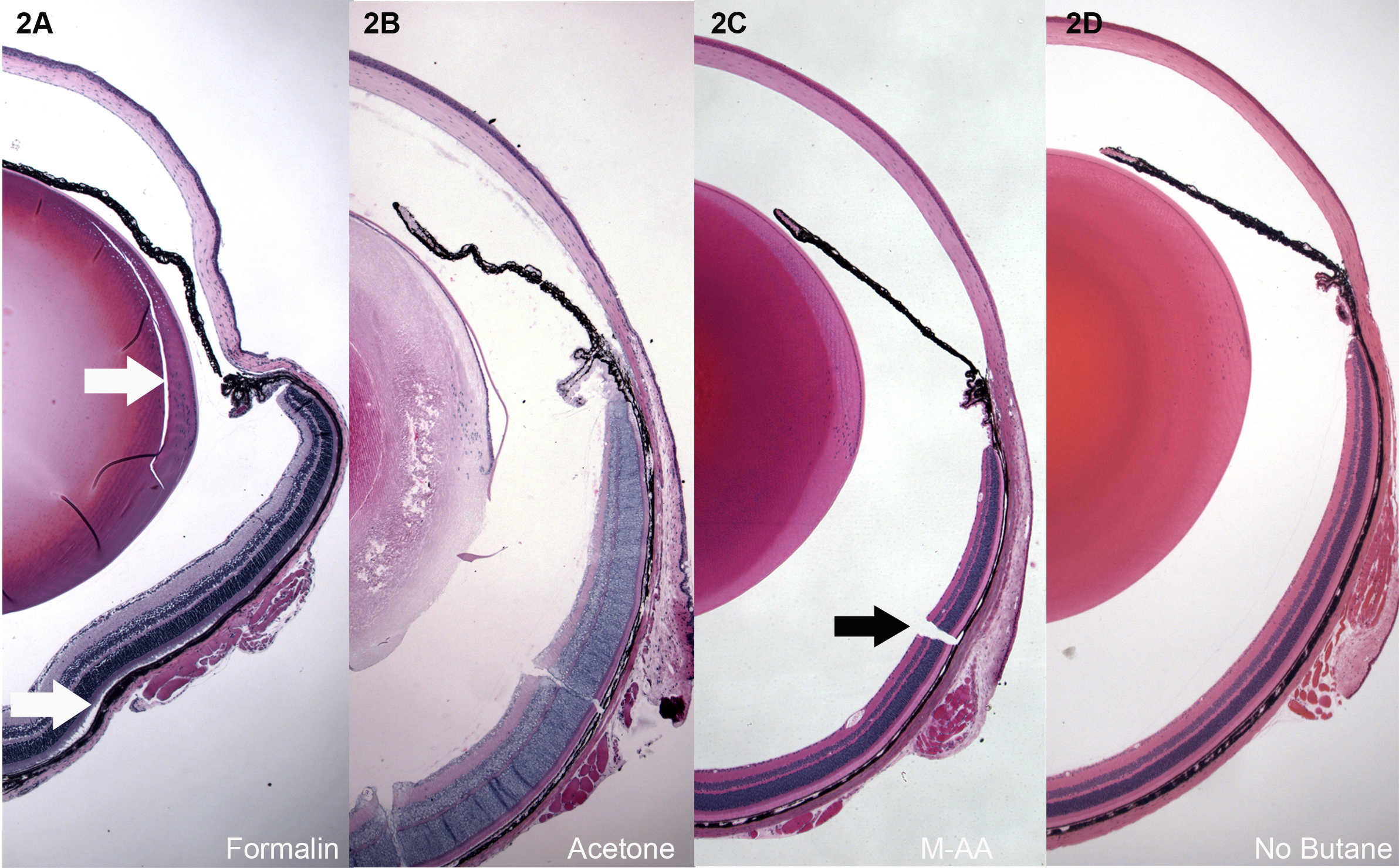
Figure 2. Low magnification overviews of H&E-stained paraffin sections of mouse eyes.
A: Immersion in 10% neutral buffered formalin for 24 h.
B: Freeze substitution in acetone.
C: Freeze substitution in 97% methanol 3% acetic acid (M-AA).
D: Same as in C, but skipping the butane freezing step, and freezing instead by direct immersion in −80 M-AA. Formalin-fixed
eyes consistently showed shrinkage and buckling, and splitting caused by shear (white arrow, lens
Figure 1A) as tissue fixed and shrank. Retinal detachment was common as well (white arrow,
A). Acetone fixation (
B) was good for the cornea, but not for most other ocular structures. Panel
C shows tissue preserved by freeze substitution in methanol, or methanol:acetic acid. This level of preservation was typical
of all the different routines described in Materials and Methods, but only the M-AA −80 is shown. Panel
D shows that good quality can be achieved without the butane step, though, as shown in
Figure 3,
Figure 4, and
Figure 5, some loss of quality can be seen on closer inspection. Higher magnification views of the retina, cornea, and lens are shown
from each of the different freeze substitution routines in subsequent images. With the exception of the not-uncommon freezing
cracks (black arrow
C), the overall relationships of ocular structures are consistently well preserved, and lacking in buckling and retinal detachment.
 Figure 2 of
Sun, Mol Vis 2015; 21:428-442.
Figure 2 of
Sun, Mol Vis 2015; 21:428-442.