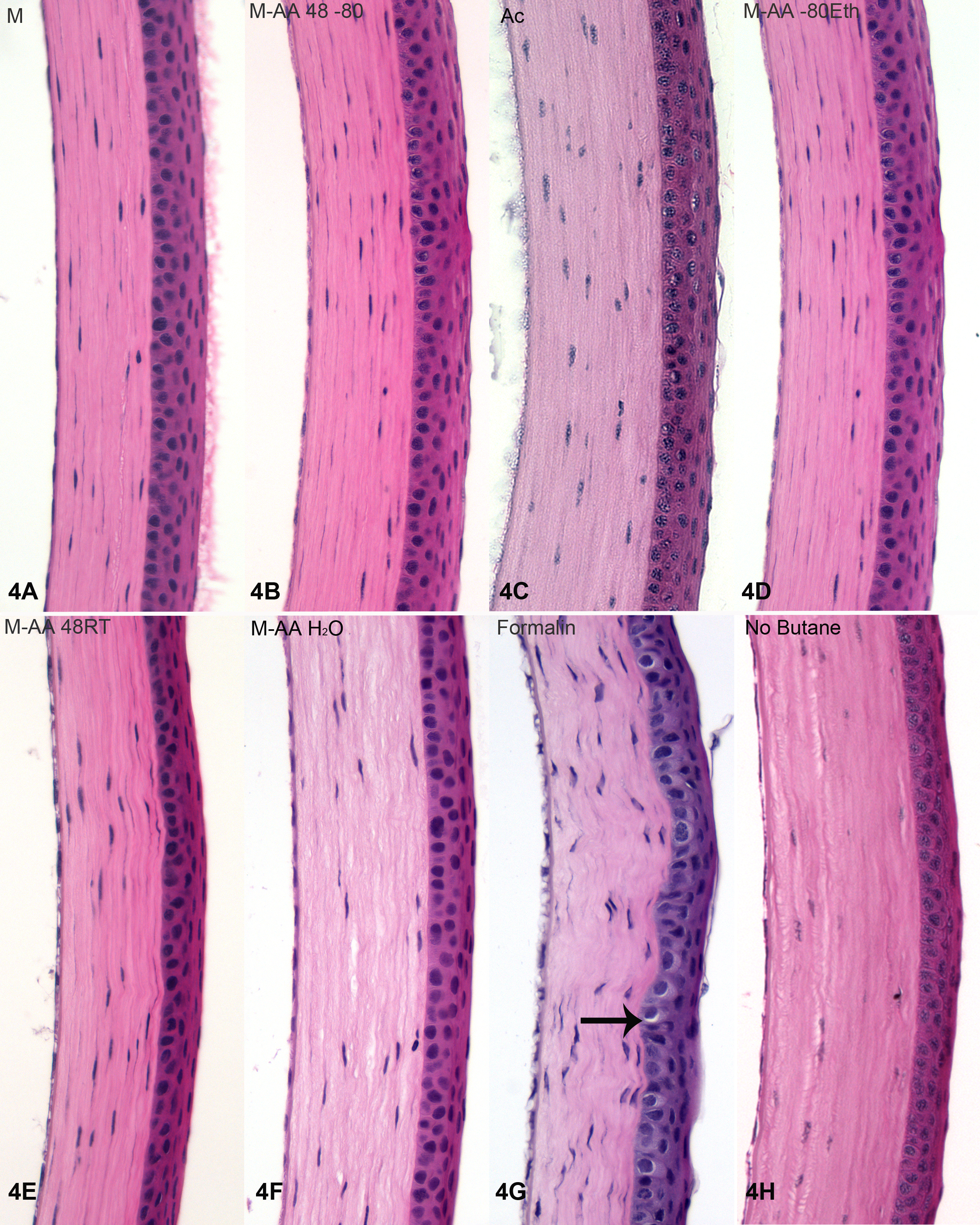
Figure 4. H&E stained corneas. Examples from each fixation routine are shown, labeled according to the key in Materials and Methods,
and as in
Figure 3. Acetone preservation was reasonably good for the cornea (
C). The shrinkage and distortion caused by formalin (
G) is evident as a nonlinear interface between the corneal epithelium and stroma. Also common to formalin fixation is perinuclear
shrinkage (arrow
G). In the rehydrated tissue (
F), there is some evidence of separation in the corneal stroma. As this was not seen in either the retina (
Figure 3F) or lens (
Figure 5F), this likely results from the presence of a strongly anionic and hygroscopic matrix that is uniquely high in the cornea
and absent from the other tissues.
 Figure 4 of
Sun, Mol Vis 2015; 21:428-442.
Figure 4 of
Sun, Mol Vis 2015; 21:428-442.