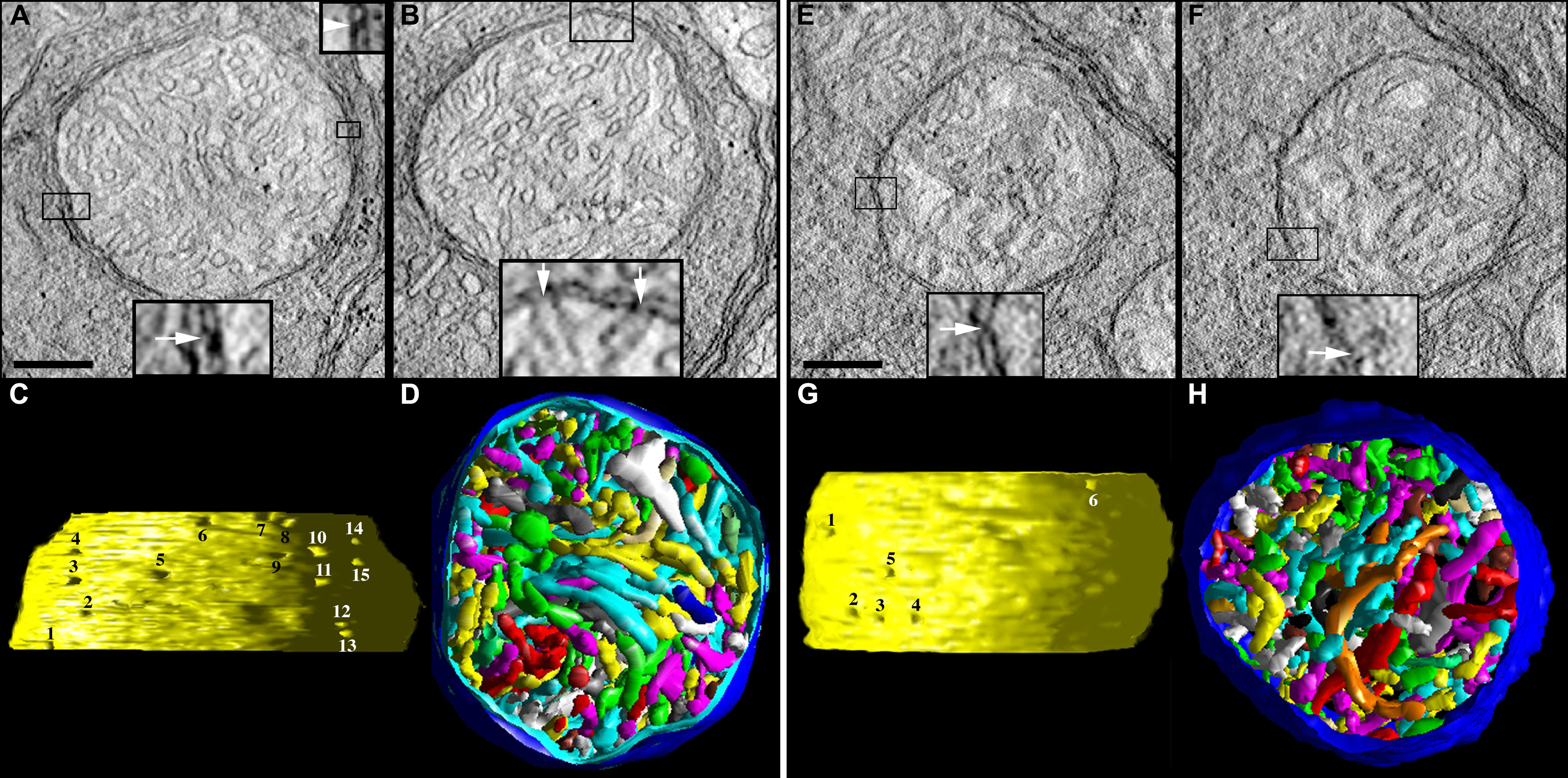
Figure 2. Control spherule and pedicle mitochondria possess many cristae, cristae with tubular and lamellar segments, narrow crista
junctions, and punctate contact sites.
A-
D: Tomographic reconstructions of a rod spherule mitochondrion from an adult control mouse are presented.
A: A 2.2 nm slice through the center of a tomographic volume of a spherule shows the membrane profiles and associations including
those of the outer and inner boundary membranes and cristae in a large mitochondrion with many cristae. The inset at the bottom
provides an example of a classic contact site (boxed and enlarged 3X), defined as the site where the outer mitochondrial membrane
(OMM) and inner boundary membrane (IBM) are joined by pinching together (arrow). The inset at the top provides an example
of a bridge contact site (boxed and enlarged 3X), defined as the site where the OMM and IBM are joined by a bridge or tether
(arrowhead). Scale bars=200 nm.
B: Another slice through the volume showing a crista junction (boxed), which is an opening, often tubular, connecting the intracristal
space with the intermembrane space. The inset at the bottom shows the opening (arrows) of two adjacent crista junctions enlarged
3X.
C: Side view of the inner membrane of the segmented and surface-rendered volume displayed with left lighting showing the size
and density of crista junctions. Crista junction openings were found to be invariably narrow, tubular, and remarkably uniform
in diameter. There are 15 numbered crista junction openings in this view.
D: Top view of the segmented and surface-rendered volume showing the outer membrane (blue) and the entire complement of 170
cristae (various colors) provides a three-dimensional feel for the packing arrangement and density of the cristae. Most of
the cristae are tubular. However, some cristae are completely lamellar or possess lamellar compartments attached to tubular
compartments, and still connect to the intermembrane space via tubular crista junctions. Compare with
Figure 7.
E–
H: Tomographic reconstructions of a cone pedicle mitochondrion from an adult control mouse are presented.
E: A 2.2 nm slice through the center of a tomographic volume of a pedicle shows a medium-sized mitochondrion. The classic contact
sites have the same structure as those in the spherule mitochondria; an example contact site is boxed and shown enlarged 3X
in the inset.
F: Another slice through the volume showing a crista junction (boxed), enlarged 3X in the inset.
G: Side view of the inner membrane of the segmented and surface-rendered volume. The crista junction architecture was found
to be similar to that in the spherule mitochondria. There are six numbered crista junction openings in this view.
H: Top view of the segmented volume showing the outer membrane (blue) and the entire complement of 165 cristae (various colors).
As with spherule mitochondria, most of the cristae are tubular. However, some cristae are lamellar or possess lamellar compartments.
Compare with
Figure 8.
 Figure 2 of
Perkins, Mol Vis 2012; 18:3029-3048.
Figure 2 of
Perkins, Mol Vis 2012; 18:3029-3048.