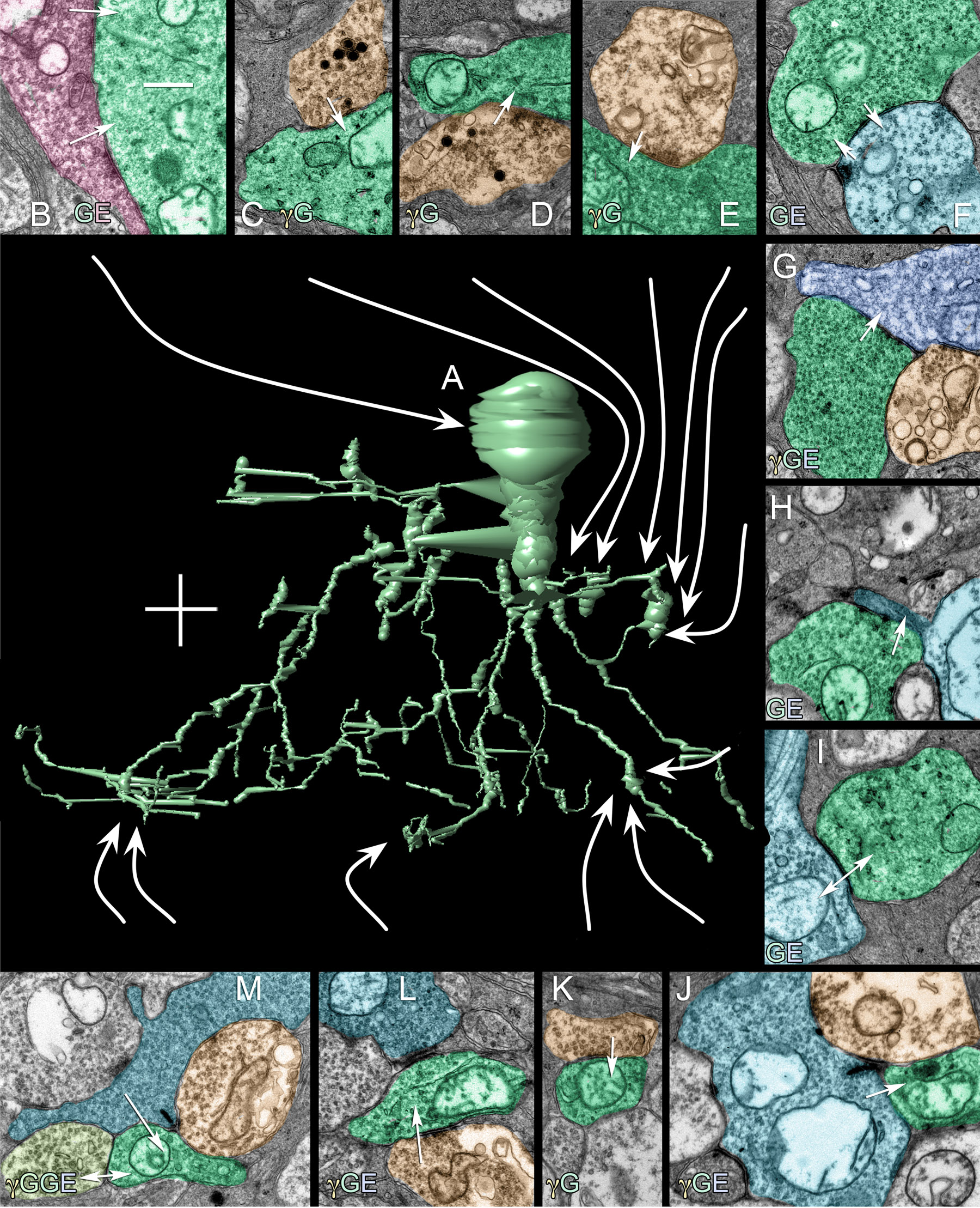
Figure 5. Numerous synaptic connections
converge on AII amacrine cell C514.
A: The central image is a
3D VikingPlot rendering of C514 (scale, 10 μm). Surrounding the cell
are instances of different synaptic connections made by C514. In each
panel, green profiles are C514, orange profiles are γ+ ACs, azure
profiles are BCs, blue profiles are GCs, red profiles are γ-, G- and
glutamate+. Arrows indicate direction of synaptic signaling and double
arrows indicate gap junctions.
B: C514 is postsynaptic to large
γ-/G- axosomatic synapses likely deriving from TH1 (tyrosine
hydroxylase immunopositive type 1) cells. However, the architecture of
the synapse is of a fast conventional transmitter, likely glutamate
(see
Figure 7
and
Figure 8).
C: C514 is postsynaptic to γ+ / peptidergic processes in the OFF
sublayer at points where dense-core, peptide vesicles form fusion
complexes.
D: C514 is postsynaptic to γ+ / peptide processes in
the OFF sublayer at a conventional inhibitory synapse.
E: C514
is postsynaptic to conventional, non-peptide γ+ processes in the OFF
sublayer.
F: C514 is both presynaptic and postsynaptic to an
OFF cone bipolar cell.
G: C514 is presynaptic to an OFF
ganglion cell.
H: C514 is presynaptic to an OFF bipolar cell.
I:
C514
is coupled to an ON cone bipolar cell.
J: C514 is
postsynaptic to an ON cone bipolar cell.
K: C514 is
postsynaptic to a γ+ amacrine cell.
L: C514 is postsynaptic to
a γ+ type AI amacrine cell.
M: C514 is postsynaptic to a rod
bipolar cell and coupled to another AII amacrine cell. The scales for
panels
B-
M are 500 nm.
 Figure 5 of Anderson, Mol Vis 2011; 17:355-379.