Figure 7. TH+ (tyrosine
hydroxylase immunopositive) cells have glutamatergic, not GABAergic
signatures. The nine panels show nine TH+ cells from a
single rabbit retina (A-I), probed for TH, glutamate and
GABA in serial 200 nm sections. Each panel shows four mappings: upper
left TH (yellow) + glutamate (blue), upper right TH (yellow) + GABA
(red), lower left glutamate alone (cyan), lower right GABA alone
(yellow). The location of each TH+ cell is circled. Each TH+
cell has a glutamate signal higher than the surrounding amacrine cell
somas and equivalent to that of a ganglion cell. TH+ cells
have no measurable GABA signal. Scale, 10 μm.
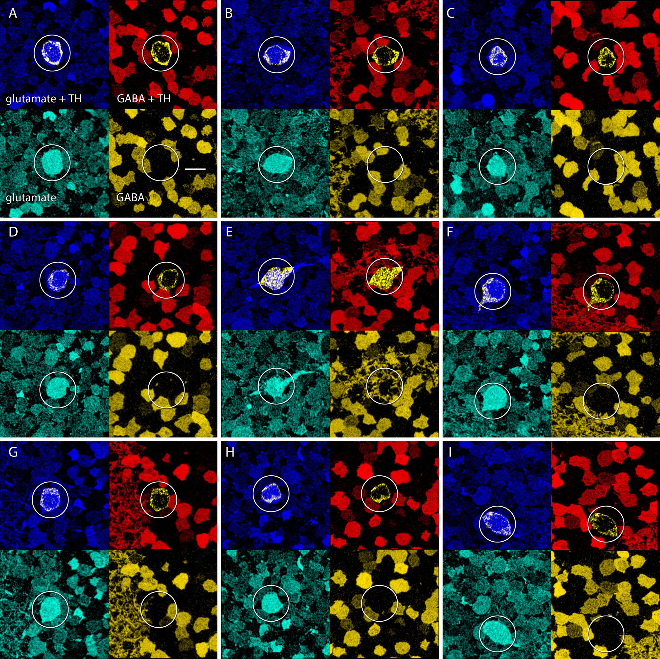
 Figure 7 of Anderson, Mol Vis 2011; 17:355-379.