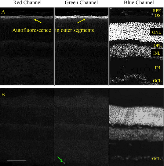
Figure 5. Representative cross-sectional micrographs show both untreated retinas and retinas two weeks after transduction with AAV2.CBA.eGFP,
but with no enzyme treatment.
A: Some sections of retina with no treatment (control) showed patchy autofluorescence in the photoreceptor outer segment layer.
This could be distinguished from GFP fluorescence because it was observed in both red and green channels (see also
Figure 6A,
Figure 7D,E).
B: Retina treated with AAV2.CBA.eGFP vector showed low transduction of the GCL. Texas Red filter is shown in the left panel;
FITC filter is shown in the center panel; DAPI filter is shown in the right panel. Calibration bar 50 μm. Ganglion cell layer
(GCL), inner plexiform layer (IPL), inner nuclear layer (INL), outer plexiform layer (OPL), Outer nuclear layer (ONL), Outer
segments (OS) and Retinal pigment epithelium (RPE). Green arrow shows a retinal ganglion cell.
 Figure 5 of
Cehajic-Kapetanovic, Mol Vis 2011; 17:1771-1783.
Figure 5 of
Cehajic-Kapetanovic, Mol Vis 2011; 17:1771-1783.