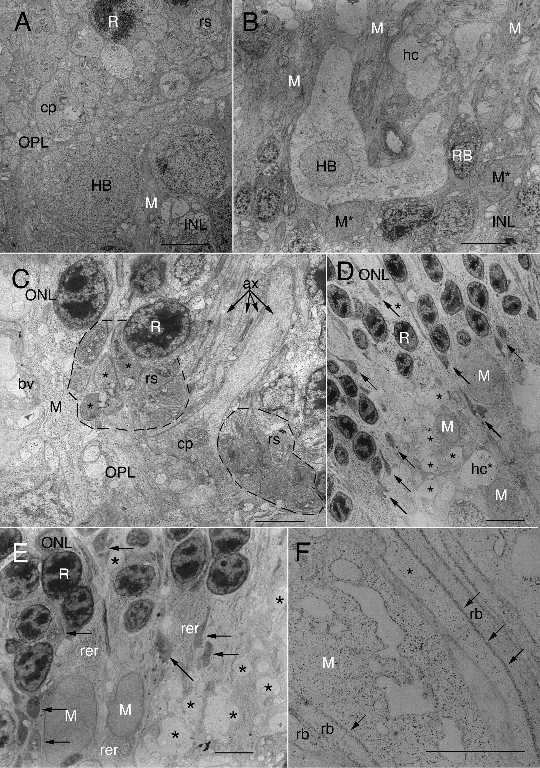
Figure 3. Electron micrographs of normal
and detached cat retina.
A: The cell body of a B-type
horizontal cell (HB) lies at the inner edge of the outer plexiform
layer (OPL). Its apical cytoplasm is densely packed with organelles.
One of its main dendritic trunks courses off to the right. Many rod
spherules (rs) are packed together in the outer OPL beneath the lowest
tier of rod cell nuclei (R). Part of a cone pedicle is shown (cp). Dark
Müller cell cytoplasm (M), though surrounding all retinal neurons, is
most evident in the inner nuclear layer (INL), where Müller cell
processes are thickest. Notice that Müller cell processes in the outer
retina are so fine as to be unresolved between neuronal processes.
Scale bar represents 10 μm.
B: In cat retina detached for 7
days, HBs appear enlarged, resulting in much lighter staining of their
cytoplasm and a decreased density of organelles. Similar
lightly-stained and enlarged profiles (hc) occur throughout the OPL.
These correspond to the ‘vacuoles’ in the OPL of
Figure 1B,C,E-G.
The OPL is disrupted lacking the typical layering of photoreceptor
terminals. Müller cell cytoplasm (M) is atypically obvious in the outer
OPL. Müller cell nuclei in the INL (M*) are less electron dense than
usual and have rounded up, losing their typical angularity. Electron
dense RB somata lie to either side of the HC, one of which is labeled.
Scale bar represents 10 μm.
C: In 7-day detached cat retina, 2
clusters (dotted outlines) of rod spherules (rs) are in their usual
position above the OPL neuropil, surrounding a cone pedicle (cp). Those
at the left have turned their basal synaptic surfaces toward one
another; 3 of them have no hilus but have apparent postsynaptic
contacts in non-invaginating or “open” configurations. (See also
Figure 4C.)
On the far left, a thick column of Müller cell cytoplasm has replaced
lost photoreceptor terminals. The axons of rods and cones (ax) are
indicated by arrows. Scale bar indicates 5 μm.
D: Ectopic
Müller cell nuclei (M) lie in the proximal ONL and OPL of 7-day
detached retina. Their processes are clearly evident in the ONL where
they surround surviving rod nuclei (R). Swollen HC processes (*) and
dendrites (hc*) are evident deep in the ONL as well as the OPL.
Electron-dense, teardrop-shaped retracting spherules (arrows) and their
axons lie at varying levels in the proximal ONL. The OPL is largely
filled with HC and Müller cell processes. Scale bar represents 10 μm.
E:
The outer OPL in a 7-day detached retina contains darkly-stained,
elongated rod spherules (arrows) that are seen at the OPL and also
within the proximal ONL amid rod nuclei (R). Large Müller cell nuclei
(M) lie within the OPL in columns of cytoplasm that often contain
extensive arrays of rough endoplasmic reticulum (rer), scattered
mitochondria, polysomes, and cytoskeletal elements. Asterisks indicate
swollen HC processes. Scale bar represents 5 μm.
F: In 7-day
detached retina, thickened rod bipolar cell dendrites (rb) and a cone
axon (*) co-fasciculate through the inner ONL with a stout Müller cell
process containing distended rER and many ribosomes. Subsurface
cross-sections of smooth endoplasmic reticulum (arrowheads) are part of
the “helical organelle” characteristic of rod bipolar cells and key to
their identification by electron microscopy. Scale bar represents 5 μm.
 Figure 3 of Linberg, Mol Vis 2009; 15:10-25.