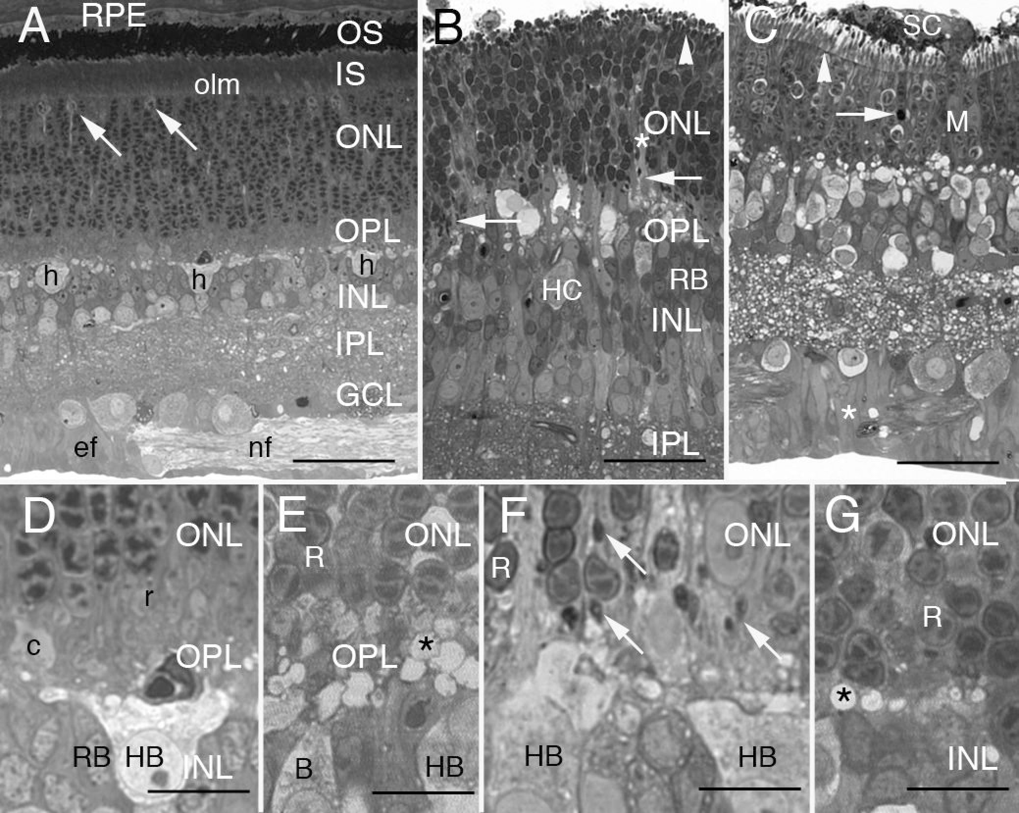
Figure 1. Light micrographs of 0.5
μm-thick resin sections of normal and detached cat retina stained with
paraphenylenediamine.
A: The layers of this radial section of
the posterior, superior temporal region of normal cat retina are
labeled to the right. Lipophilic paraphenylenediamine (PPDA) intensely
stains the photoreceptor outer segments (OS). Except for the few
lightly staining cone nuclei (arrows) just beneath the outer limiting
membrane (olm), the 11 or more ranks of nuclei comprising the outer
nuclear layer (ONL) are all rods. The outer plexiform layer (OPL)
begins where the innermost row of rod nuclei ends and spans to the line
of lightly stained horizontal cell somata, 3 of which are shown (h).
More proximal still are the inner nuclear (INL), inner plexiform (IPL),
and ganglion cell (GCL) layers. Bundles of ganglion cell axons (nf) lie
among Müller cell end feet (ef), bordering the retina’s inner limiting
membrane. An enlargement of the central region is shown in
D.
Abbreviations: retinal pigment epithelium (RPE); inner segments (IS).
Scale bar represents 40 μm.
B: In this slightly oblique section
through the posterior, superior nasal region of 7-day detached retina,
photoreceptor OS and IS have collapsed and extend just a few
micrometers above the olm (white arrowhead). Only 8 to 10 rows of rod
nuclei form the ONL, including regions of loosely packed cells (*). The
inner margin of this layer is no longer well defined. Many densely
stained retracting rod spherules lie in this region (arrows). Most of
the lightly stained profiles of varying sizes in the outer OPL are the
swollen processes of the B-type horizontal cell (HC). A B-type HC cell
body (HB) with its prominent nucleolus lies in the outermost layer of
the INL while several rod bipolar cells (RB) cluster to the right.
Scale bar represents 35 μm.
C: After 28 days of detachment, the
ONL of posterior superior temporal retina has only 5 to 7 rows of rod
nuclei including one cell shown here undergoing apoptosis (arrow).
“Vacuoles” in the OPL are actually swollen processes of the B-type HC.
Profiles of retracting spherules, so prominent at 7 days of detachment (
B,
F), are no longer obvious at 28 days. The Müller cells show
dramatic changes including enlargement of the overlapping end feet (*),
migration of their nuclei through the outer retina (M), and
participation in the formation of a subretinal scar (SC) that spreads
out over the photoreceptor layer. Arrowhead indicates olm. Scale bar
represents 35 μm.
D: The stratified OPL of normal cat retina is
shown at higher magnification. This same region is seen in the middle
of
A. From the top of the figure, the innermost 5 to 6 rows of
rod nuclei are densely packed. The outer half of the OPL contains the
photoreceptor terminals: several rows of tightly packed rod spherules
(r) lie distal to cone pedicles (c). The inner half of the OPL is the
neuropil itself. A small capillary containing a red blood cell is
transected just above the cell body of a B-type HC (HB) at the outer
margin of the INL. RB nuclei also lie in the outer half of the INL.
Scale bar represents 10 μm.
E: Three days after detachment, the
feline outer retina retains much of its typical stratification.
Although voids appear among them, rod cell bodies (R) are still closely
packed above clustered rod spherules that appear to stain less
intensely than normal, but retain their normal size. The underlying
neuropil is heavily populated with vacuole-like profiles (*) identified
as the swollen branches of hypertrophied HC dendrites and axon
telodendria. A portion of an HB shows lightly stained cortical
cytoplasm ballooning past the typically dense field of organelles lying
near the nucleus. B is an unidentified cone bipolar cell. Scale bar
represents 10 μm.
F: Disrupted organization of the inner ONL
and OPL a week after detachment. The inner ONL has loosely packed rod
nuclei (R) separated by thickened Müller cell processes. Large,
lightly-staining, ectopic Muller cell nuclei are found here as are the
small dark profiles of retracting rod spherules (arrows). Lightly
stained HCs have swollen cell bodies (HB). The largely empty cortical
cytoplasm extends beyond a diverse field of perinuclear organelles.
Pale profiles scattered throughout the OPL are swollen HC processes.
Scale bar represents 10 μm.
G: Almost a month after detachment,
the cell bodies of surviving rods (R) in the ONL directly front on a
narrowed OPL neuropil that contains vacuole-like cross-sections of
hypertrophied HC processes (*). Though not evident at this
magnification, scattered spherules can still be identified by electron
microscopy (
Figure
4I). Scale bar represents 10 μm.
 Figure 1 of Linberg, Mol Vis 2009; 15:10-25.