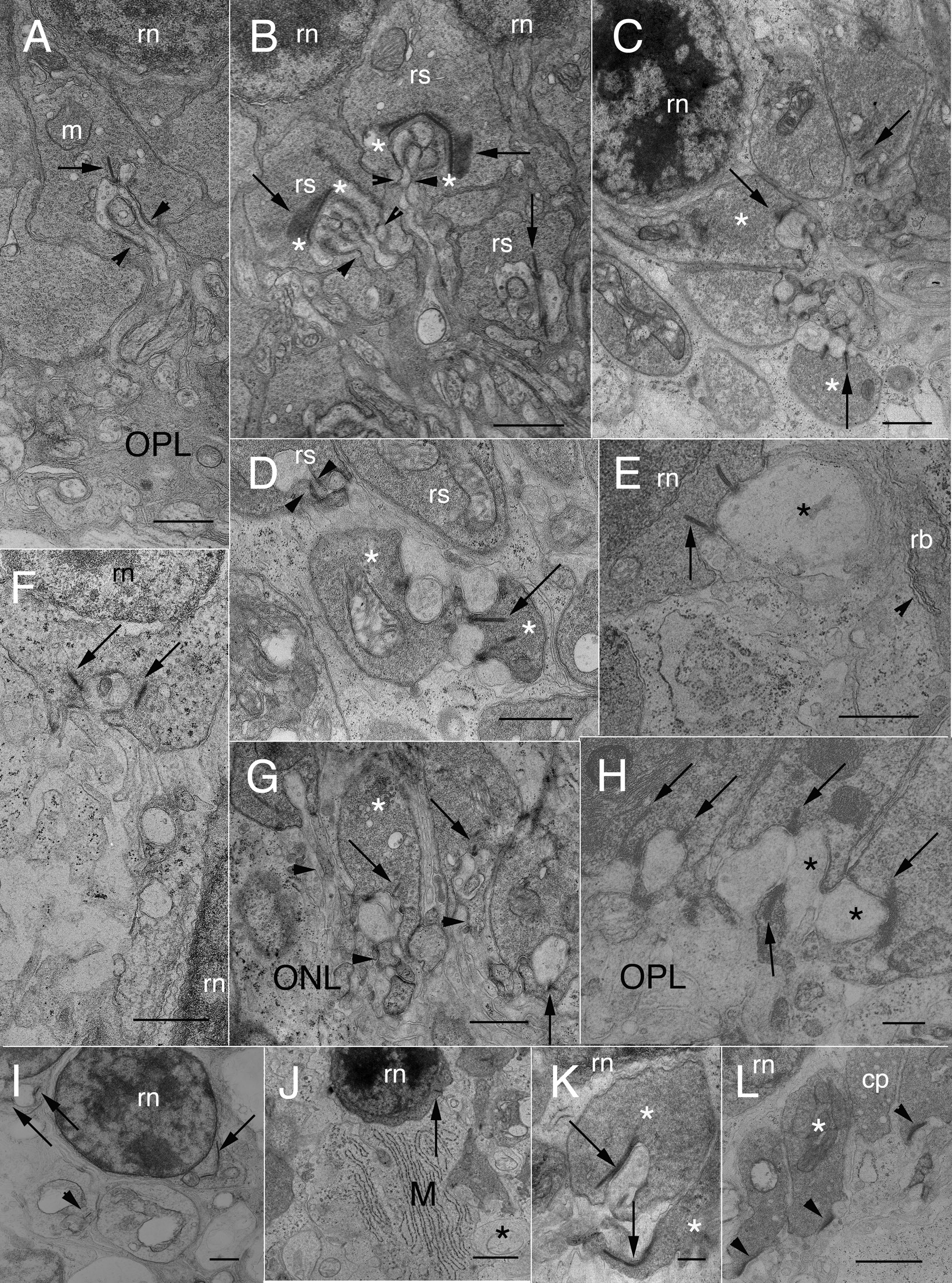
Figure 4. Electron micrographs of normal
and detached cat retina.
A: A rod spherule lies between the OPL
neuropil and the innermost tier of rod cell nuclei (rn). The
presynaptic cytoplasm is crowded with a uniform population of 35–45 μm
light cored synaptic vesicles. A synaptic ribbon (arrows) opposes fine,
invaginating HCat telodendria and rod bipolar dendrites. The
innervating processes pass through the hilus (arrowheads) to enter the
invagination. Mitochondria (m) typically lie near the synapse. Scale
bar represents 0.8 μm.
B: In normal cat retina, 3 neighboring
rod spherules (rs) show details of presynaptic and postsynaptic
architecture. In two rod spherules, the synaptic ribbon (arrows) is
seen chiefly en face. In addition to the dense presynaptic populations
of synaptic vesicles, the upper spherule also contains profiles of ser
cisternae or endosomes, two of which lie near a mitochondrion. At the
base of the synaptic ribbons, asterisks (*) demarcate the ends of the
particularly electron-dense arciform densities. HCat telodendria and
rod bipolar (RB) dendrites pass through a basal hilus to enter their
respective synaptic invaginations (arrowheads). The rod nucleus is
labeled (rn). Scale bar represents 0.8 μm.
C: This area of the
OPL in 7 day detached cat retina is enlarged from
Figure 3C.
Although these spherules lie in their usual location and show little
evidence of retraction, their ultrastructure is abnormal. The spherules
themselves are turned in such a way that their basal synaptic surfaces,
instead of facing the OPL, face each other around a cluster of
postsynaptic processes. Two of the spherules (*) have shallow
invaginations without evidence of any hilus. Swollen HCat telodendria
contact these spherules in “open” configurations. Synaptic ribbons
(arrows) are seen in three spherules. All the spherules in this
location are filled with synaptic vesicles. Interestingly, the two
profiles of a synaptic ribbon in the bottom spherule have arciform
densities that appear to project as synaptic ridges between apposing
horizontal cell (HC) lateral elements. Scale bar represents 0.75 μm.
D:
Several retracting rod spherules in 7-day detached cat retina lie in
the lower outer nuclear layer (ONL) enveloped by Müller cell cytoplasm.
The uppermost spherule is invaginated; one HC lobe is visible as is the
hilus (arrowheads) through which the postsynaptic processes pass. Two
subjacent spherules (*) make open contacts with swollen HC axon
telodendria. One contains a synaptic ribbon (arrow) that shares a HC
lobe with a neighboring ribbon. Both ribbons appear to extend from
arciform densities that project outward as synaptic ridges. Spherules
above and below them lack synaptic ribbons in this plane of section,
but have multiple mitochondria. Synaptic vesicles populate all of the
terminals. Scale bar represents 0.8 μm.
E: A section through
basal perinuclear cytoplasm of a rod soma in the mid-ONL of 7-day
detached cat retina shows numerous synaptic vesicles and two
cross-sections of synaptic ribbons. One to the left is presynaptic to
two vesicle-containing HC processes, one of which (*) is very swollen
and also postsynaptic to the other ribbon, both in open,
non-invaginated configurations. Subjacent Müller cell cytoplasm has
distended rer cisternae and numerous polysomes. Scale bar represents
0.8 μm.
F: Two rod nuclei (rn) lie in the mid- to lower ONL of
cat retina detached for 7 days. The shallow perinuclear synaptic
invagination of the upper cell body contains two pale lateral elements
and a small, dark central element. Arrows point to cross-sections of
synaptic ribbons (perhaps the same one) that lack arciform densities
and have only a few synaptic vesicles lying near them. Presynaptic
cytoplasm also contains a few endosomes and scattered polysomes. Scale
bar represents 0.6 μm.
G: Four partially retracted spherules
lie in the inner ONL of a 7-day detached retina. All have their
synaptic surfaces facing the inner retina. These terminals are filled
with synaptic vesicles and have synaptic ribbons (arrows) presynaptic
to electron-lucent HCat telodendria. One of the spherules (*) makes an
open contact with postsynaptic processes and lacks the normal synaptic
invagination. RB outgrowths (arrowheads) lie near these synapses but do
not enter the invaginations. Scale bar represents 0.8 μm.
H:
Three rod spherules in 7-day detached cat retina remain at the OPL but
nevertheless lack typical synaptic invaginations. Presynaptic cytoplasm
is crowded with synaptic vesicles. Synaptic ribbons (arrows) appose
swollen and electron-lucent HCat telodendria in open configurations.
The HCat processes innervating two adjacent spherules (*) directly
appose each other. Scale bar represents 0.3 μm.
I: Two rod
nuclei (rn) in 28-day detached cat retina front on the OPL. Both
contain a perinuclear basal invagination with synaptic ribbons
(arrows). Subjacent spherules lie in their normal position; one also
contains a synaptic ribbon (arrowhead). The density of the synaptic
vesicle population is abnormally low. Scale bar represents 0.6 μm.
J:
Low power survey of the inner ONL in cat retina detached for 7 days.
Several retracting rod spherules are loosely packed along with a rod
soma (rn) containing a cross-section of an indistinct synaptic ribbon
(arrow) that apposes an HCat process without evidence of any
invagination, arciform density, or RB dendritic contact. Only two or
three synaptic vesicles associate with the ribbon in this plane of
section. Müller cell cytoplasm (M) enshrouding these structures
contains prominent arrays of parallel rer cisternae. Asterisk (*)
indicates swollen HCat process. Scale bar represents 1.5 μm.
K:
Two retracting spherules (*) in cat retina detached for 7 days contain
synaptic ribbons (arrows) that lie almost parallel to their presynaptic
membranes. Arciform densities are not discernible. The upper spherule
is invaginated, the lower is not. Both are crowded with synaptic
vesicles. Post-synaptic HC lateral elements contain flattened light
cored vesicles of various sizes. Scale bar represents 0.3 μm.
L:
In 7-day detached cat retina, three retracting rod spherules in the
lower ONL are surrounded by Müller cell cytoplasm (M). All three show
basal densifications (arrowheads) but no ribbons or other structural
characteristics of rod synapses. The presynaptic cytoplasm contains
vesicles, one or more mitochondria (*), endosomes, and scattered
polysomes. The edge of a cone pedicle (cp) is seen. Scale bar
represents 1.2 μm.
 Figure 4 of Linberg, Mol Vis 2009; 15:10-25.