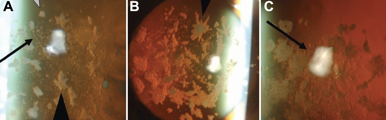
Figure 3. Lattice lines in patient's
cornea. The lattice lines referred to in
Figure 2 are better visualized in
the higher magnifications of
Figure 2C,
Figure 2F,
and
Figure 2I
(
Figure 3A-C,
respectively). The lattice lines are easily seen in
A and
C
(black arrows), but not in
B.
 Figure 3 of Yamada, Mol Vis 2009; 15:974-979.