Figure 2. Slit-lamp photographs of the
proband and her two sisters. Slit-lamp photographs of the right eye of
II-1 (A–C), the left eye of II-2 (D–F), and
the left eye of II-3 (G–I) are shown. Granular deposits
(gray arrowheads) and star-shaped deposits (black arrowheads) were
observed in all three patients (C,F,I) whereas
thin lattice lines (black arrows) were observed only in II-1 (C)
and II-3 (I). Nodular deposits were apparent mostly in the
superficial-to-middle portion of the corneal stroma in all three
patients (B,E,H).
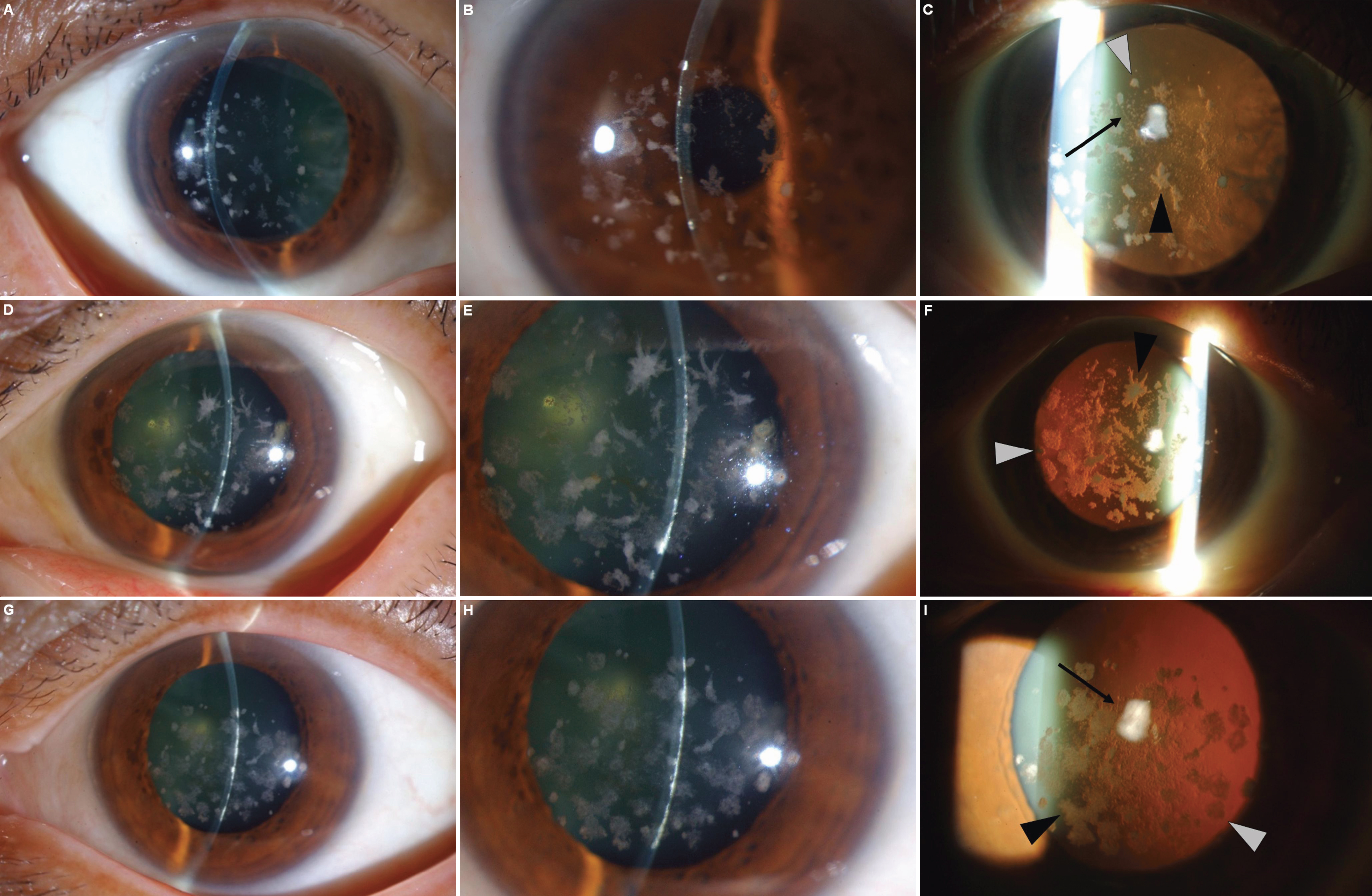
 Figure 2 of Yamada, Mol Vis 2009; 15:974-979.