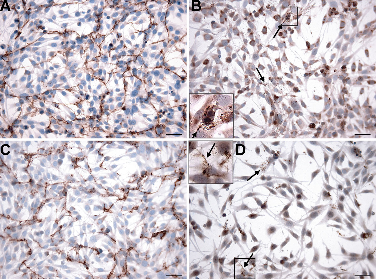
Figure 4. Immunocytochemical analyses of cultured Müller cells in medium with G5. Types I (
A) and V (
C) collagen show clear extracellular fibrillar threads and less intracellular staining compared to
Figures 2A and
2E, respectively. Types II (
B) and XI (
D) collagen show some fine extracellular threads and small granules (arrows) and decreased intracellular staining compared
to
Figures 2B and
2I, respectively. In the inlays of
Figures 4B and
4D, the extracellular collagen is magnified two times. Bars in each panel equal 50 μm.
 Figure 4 of
Ponsioen, Mol Vis 2008; 14:652-660.
Figure 4 of
Ponsioen, Mol Vis 2008; 14:652-660.