Figure 2. Immunocytochemical analyses of cultured Müller cells in medium with fetal bovine serum. A: Type I collagen shows a granular staining with a variable intensity between cells. B: Type II collagen is seen as a faint staining in the cytoplasm. C: Type III collagen is positive in all cells. D: Type IV collagen is visible as a strong granular cytoplasmic staining. E: Type V collagen shows mainly staining in the cytoplasm. F: In the case of type VI collagen, the cells are predominantly stained in the cytoplasm. G: Type VII collagen is faintly positive in the cytoplasm. H: Type IX collagen is also present in the cytoplasm. I: Type XI collagen is primarily seen in the cytoplasm. Bars in all panels equal 50 μm.
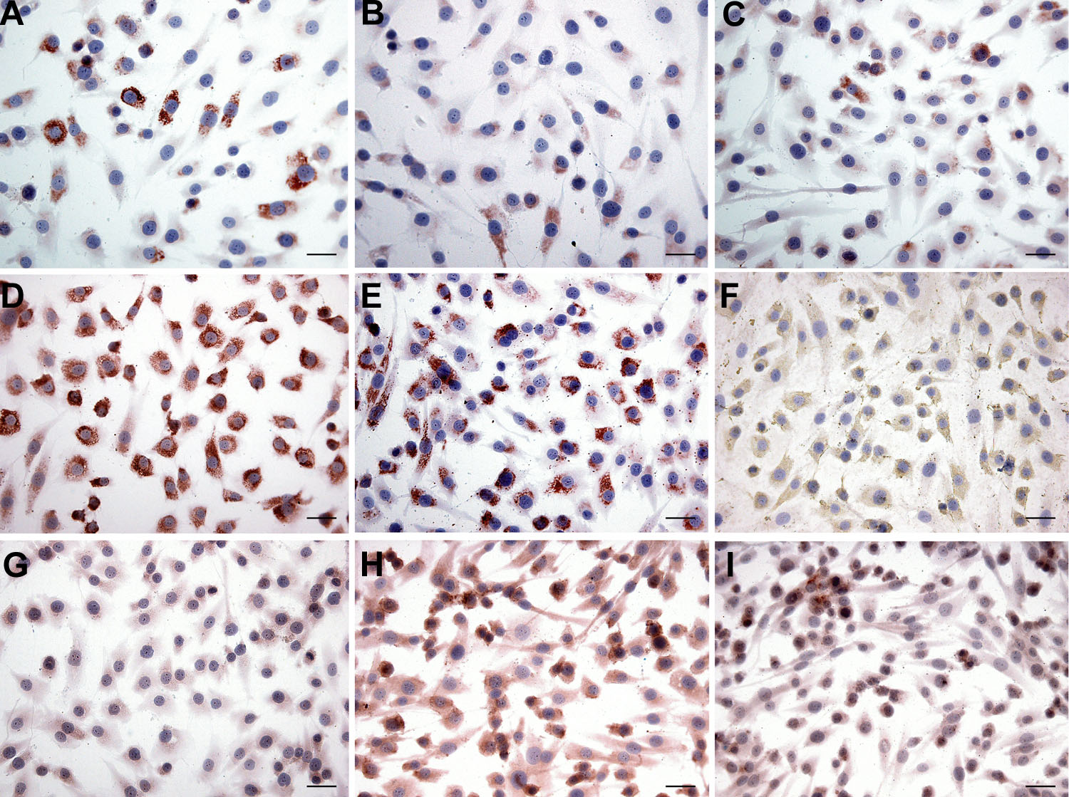
 Figure 2 of
Ponsioen, Mol Vis 2008; 14:652-660.
Figure 2 of
Ponsioen, Mol Vis 2008; 14:652-660.