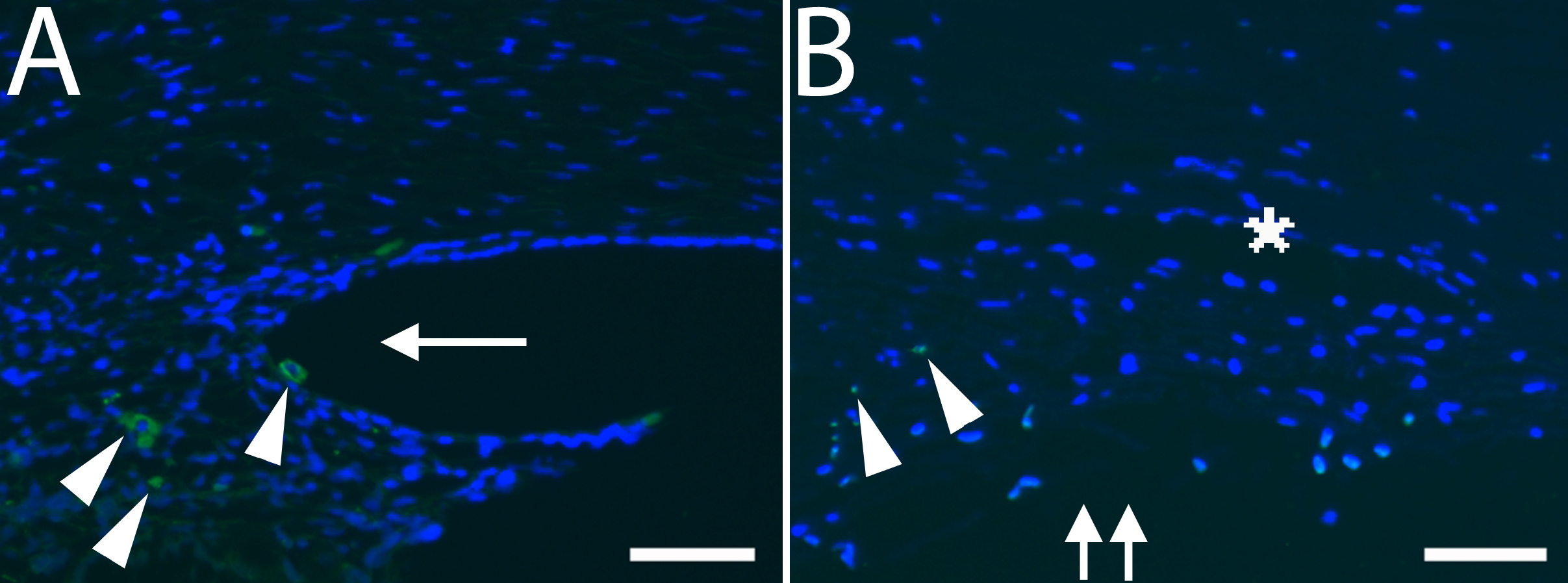
Figure 3. Immunohistochemistry and TUNEL
assay.
A: Magnified image of CD68 immunostaining at F15w from
Figure 2.
Note the cytoplasmic staining of cells in the developing ciliary body
(arrowhead). The other green stain within the nucleus above is
artifact. F, fetus; w, week. An arrow indicates an iridocorneal angle.
Double arrows indicate the trabecular meshwork. Scale bar=50 μm.
B:
Magnified image of TUNEL staining in 48-year-old adult eye from
Figure 4.
Note the TUNEL positive nuclei that appear double stained with DAPI
(arrowhead) in the trabecular meshwork, which is indicated by an
asterisk. Double arrows indicate the trabecular meshwork. Scale bar=50
μm.
 Figure 3 of Meghpara, Mol Vis 2008; 14:2492-2498.