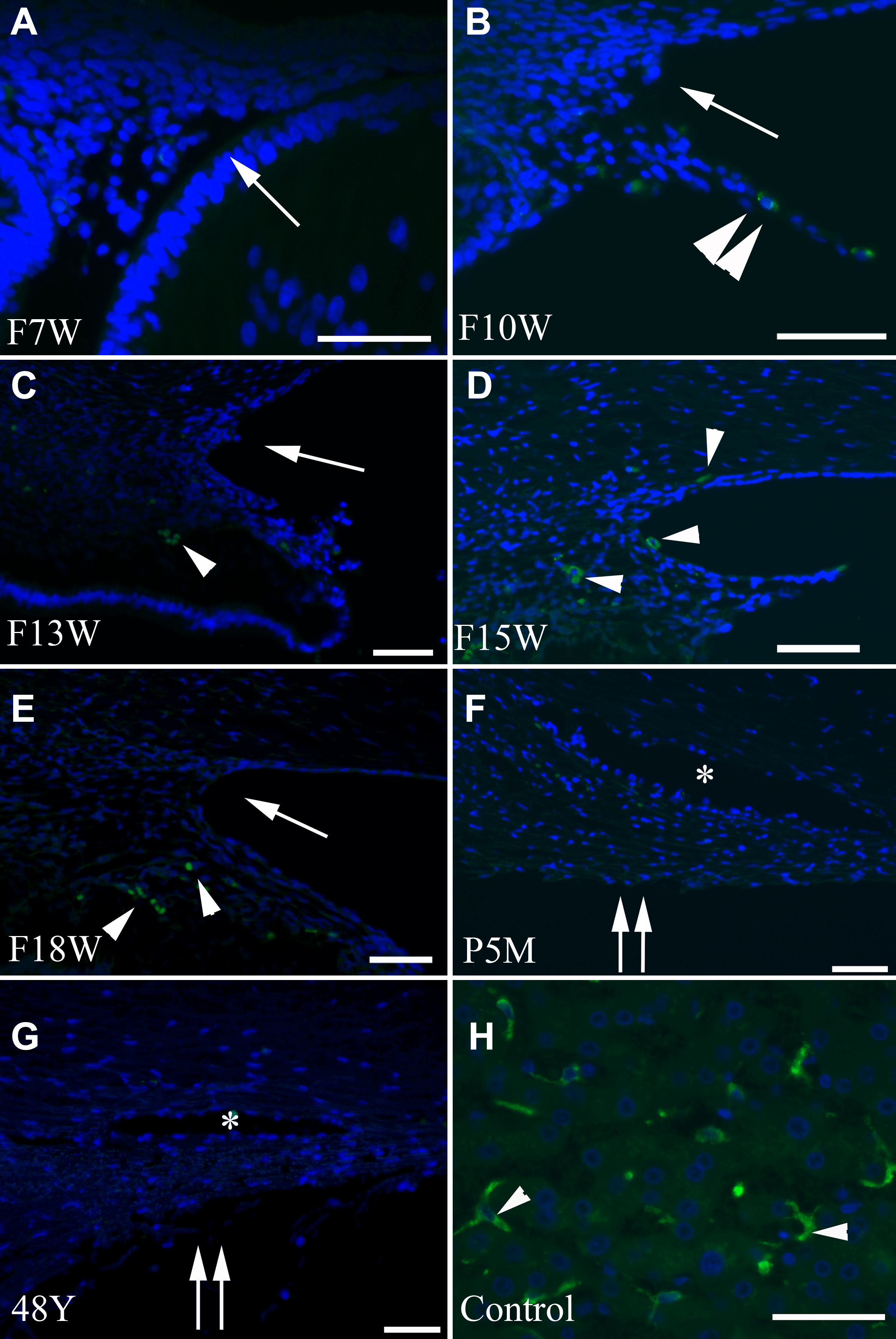
Figure 2. Photomicrograph panel showing
CD68 immunostaining visualized with DTAF and nuclei counterstained with
DAPI. The developing trabecular meshwork and surrounding areas do not
show CD68 positive cells (
A–
G). Few CD68 positive cells,
labeled in green, were observed in the iris stroma and anterior ciliary
body from F10w through F18w (
B–
E; arrowhead) and absent
at later stages (
F and
G). Note positive CD68 staining (
H)
in the human liver dendritic cells (arrowhead). F, fetus; w, week; m,
month; y, year; P: postnatal. The asterisk indicates the location of
Schlemm’s canal. An arrow indicates an iridocorneal angle. Double
arrows indicate the trabecular meshwork. Scale bar=50 μm. Panel
D
is enlarged in
Figure
3A to clearly show positively stained macrophages. Eyes from
other fetal or adult tissues/stages that are not shown in the panel did
not show CD68 positive macrophages.
 Figure 2 of Meghpara, Mol Vis 2008; 14:2492-2498.