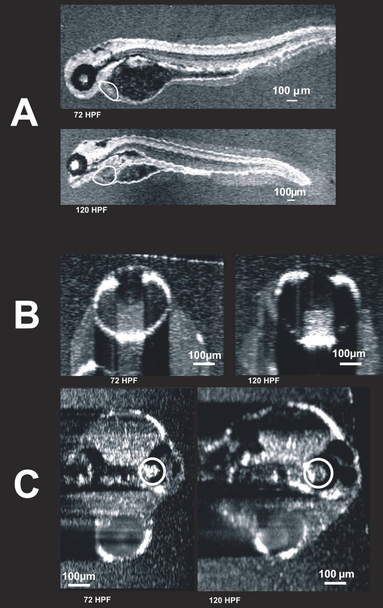
Figure 10. Visualization of individual
animals imaged on two different days. These images were obtained from
the same embryo on two different days: 72 hpf and 120 hpf. C-mode
images of the heart (
A, circled), eye (
B), and ear (
C,
circled) are presented. The heart is also visible in
C, but
blurred due to averaging over multiple cardiac cycles. It is possible
that the first imaging session altered development. To compare the 120
hpf twice-imaged embryos to 120 hpf embryos imaged only once, refer to
Figure 6
and
Figure 9.
 Figure 10 of Kagemann, Mol Vis 2008; 14:2157-2170.