Figure 6. Visualization of developing
internal anatomy of zebrafish embryos. Twenty-four hours post
fertilization (hpf): A C-mode slice (left, 750 μm) centered on the eye,
and a repeated line scan (right, 750 μm) centered on the brain and eye
provide visualization of numerous internal structures within the brain,
eye, and gut, as well as fluid spaces developing as ventricles within
the brain. 48 hpf: C-mode slices at the level of the ear (left, 750 μm)
and notochord (right, 1.5 mm). Microscopic structures of the eye,
brain, gut, and heart can be visualized throughout. Blood within
vessels and the heart create a bright reflection when isolated in a
sagittal plane (left) while casting shadows on underlying tissues
(right). 72 hpf: An averaged repeated line scan of the eye (left, 750
μm) reveals the cornea, lens, retina, and retinal pigment epithelial
and choriocapillaris complex (RPE/CC) layer of the right eye of a
zebrafish embryo. The RPE/CC of the left eye is the only structure
within the left eye with sufficient reflectance to be observed. The
C-mode slice (right, 1.5 mm) centered on the heart provides
visualization of both chambers of the heart as well as numerous
structures within the gut and brain. 120 hpf: An averaged repeated line
scan of the eye (left, 750 μm) shows the cornea, lens, retina, and
RPE/CC layer of the right eye. A C-mode slice (right, 4 mm) displays
structures of the heart, gut, and brain, documenting the development
that has occurred in only 120 hpf.
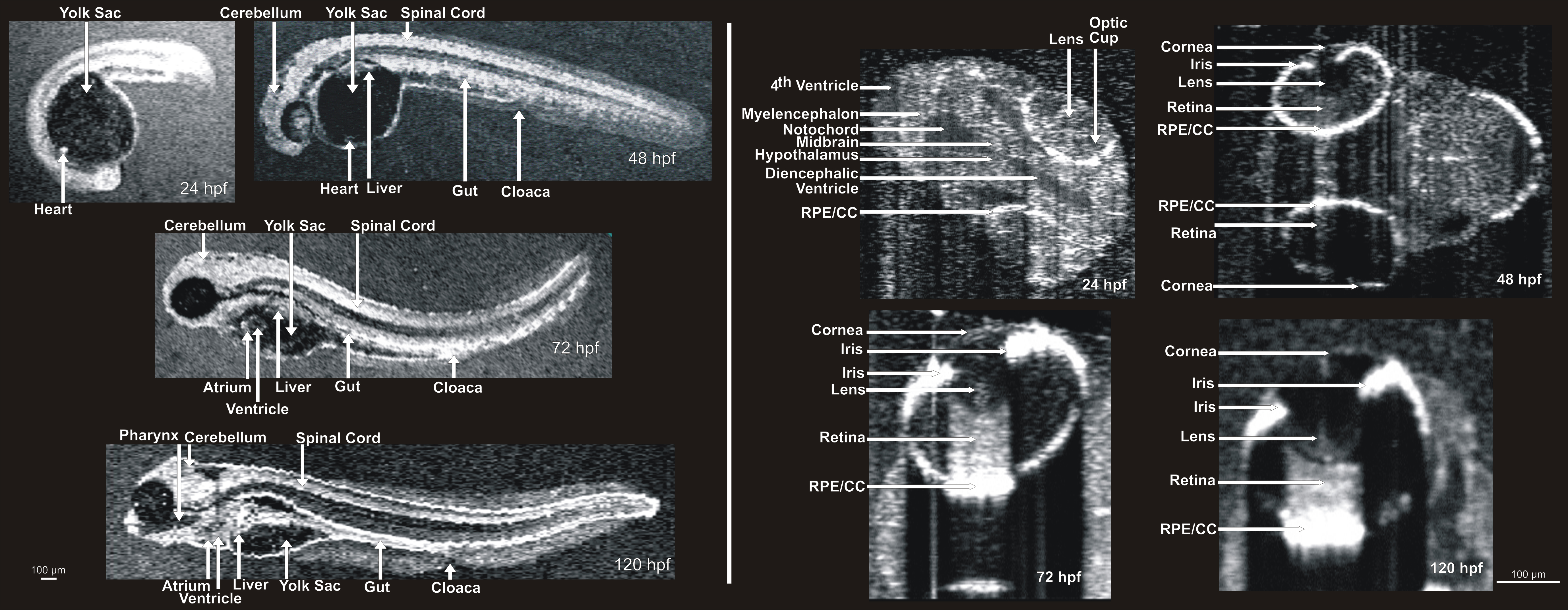
 Figure 6 of Kagemann, Mol Vis 2008; 14:2157-2170.