Figure 9. Side to side comparison of head
and body structures visualized by SD-OCT and histology. SD-OCT images
(left in each pair) were obtained noninvasively from living embryos,
leaving them healthy and capable of continued growth. Similar
structural data was obtained histologically, requiring sacrifice and
sectioning before any information was obtained, and guaranteeing that
any interesting structural observations can never be followed
longitudinally. A shows the development of the eyes and
ventricle of a 24 hours post fertilization (hpf) embryo. B and C
show development of the spine and eyes of a 48 hpf embryo respectively.
D and E show the development of the eye, spine, and
liver at 72 hpf. F, G, and H show the
development of the eye, ear, heart, and spine of the 120 hpf embryo.
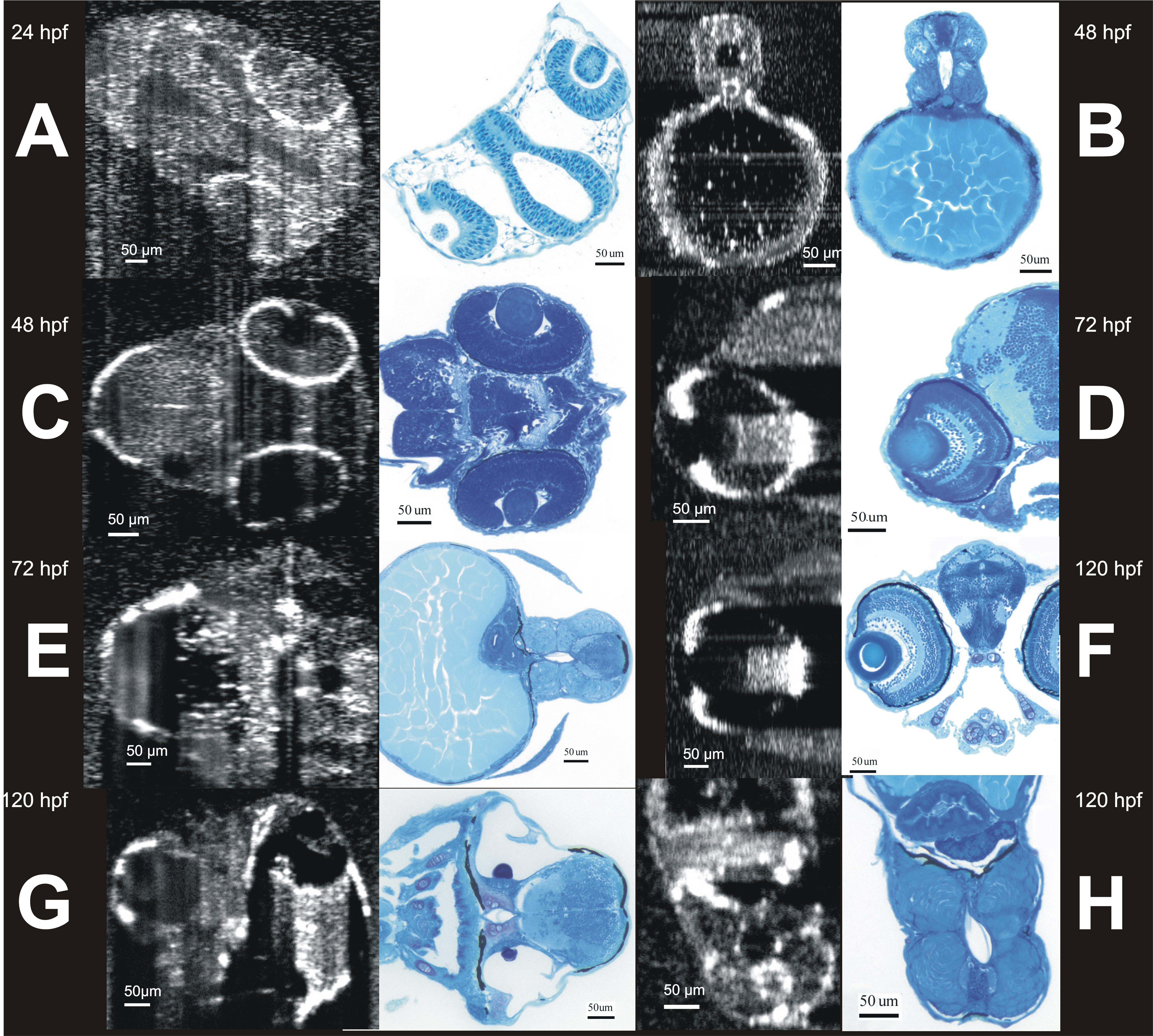
 Figure 9 of Kagemann, Mol Vis 2008; 14:2157-2170.