![]() Figure 2 of
Wilkinson-Berka, Mol Vis 2007;
13:1529-1538.
Figure 2 of
Wilkinson-Berka, Mol Vis 2007;
13:1529-1538.
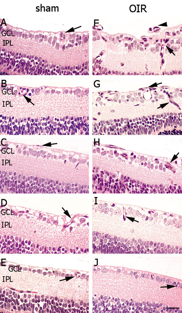
Figure 2. Three μm paraffin sections of inner retina from mice with oxygen induced retinopathy and treated with mouse GHr ASO, ATL 227446, using an early intervention protocol
Oxygen induced retinopathy is associated with pathological angiogenesis in the inner retina compared to sham mice, which is reduced with ATL 227446. The following abbreviations are used: oxygen induced retinopathy (OIR); growth hormone receptor (GHR); antisense oligonucleotide (ASO); ganglion cell layer (GCL); inner plexiform layer (IPL) and blood vessel profiles (BVPs). The sections are stained with hematoxylin and eosin. Magnification x150. Scale bar equals 50 μm. A is sham+vehicle, B is sham+5 mg/kg ATL 227446, C is sham+10 mg/kg ATL 227446, D is sham+20 mg/kg ATL 227446, E is sham+30 mg/kg ATL 227446, F is OIR+vehicle, G is OIR+5 mg/kg ATL 227446, H is OIR+10 mg/kg ATL 227446, I is OIR+20 mg/kg ATL 227446, and J is OIR+30 mg/kg ATL 227446. Retina in all sham groups (A to E) appeared normal with BVPs (arrows) in the inner retina. In OIR vehicle controls (F), BVPs (arrows) were present in the inner retina and also adherent to the retinal surface (arrowhead). In OIR, 5 mg/kg ATL 227446 (G) did not alter BVPs, however higher doses reduced BVPs (H, I, J) to a greater extent than late intervention (Figure 1 and Figure 2).