![]() Figure 6 of
Cameron, Mol Vis 2005;
11:775-791.
Figure 6 of
Cameron, Mol Vis 2005;
11:775-791.
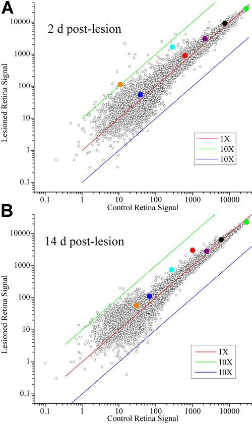
Figure 6. Lesion-induced changes in zebrafish retinal gene expression
Transcript levels are plotted for the 2 d (A) and 14 d (B) post-lesion conditions. The samples of post-lesion retinas are compared to their animal-matched control retina samples. Representative genes are indicated by the large circles, color coded to match the qPCR data plotted on Figure 9 and Figure 11: green, rhodopsin; black, pgi1; purple, cdh2; red, γ-tubulin; cyan, GAP43; blue, plasticin; orange, cxcr4b.