![]() Figure 3 of
Cailleau, Mol Vis 2005;
11:472-481.
Figure 3 of
Cailleau, Mol Vis 2005;
11:472-481.
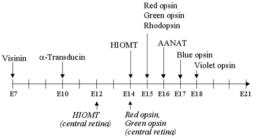
Figure 3. Schematic representation of gene activation steps in the developing chicken retina
This graph summarizes the data presented in Figure 1 and Figure 2. On a time scale spanning embryonic day 7 (E7) to E21 (hatching), arrows point to the dates of appearance of the indicated mRNAs, as detected by northern blot analysis. The dates refer to analyses performed on total retina, except when otherwise indicated (italics).