![]() Figure 5 of
Tasheva, Mol Vis 2004;
10:544-554.
Figure 5 of
Tasheva, Mol Vis 2004;
10:544-554.
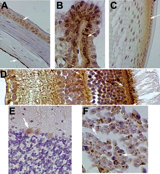
Figure 5. Higher magnification IHC micrographs of selected mouse tissues
Some mouse tissues from Figure 4 and Figure 6 were examined at higher magnification. Positive immunostaining appears brown and is highlighted by arrows. A Cornea. B: Ciliary body. C: Lens. D: Retina. E: Cerebellum. F: Peripheral nerve.