![]() Figure 4 of
Tasheva, Mol Vis 2004;
10:544-554.
Figure 4 of
Tasheva, Mol Vis 2004;
10:544-554.
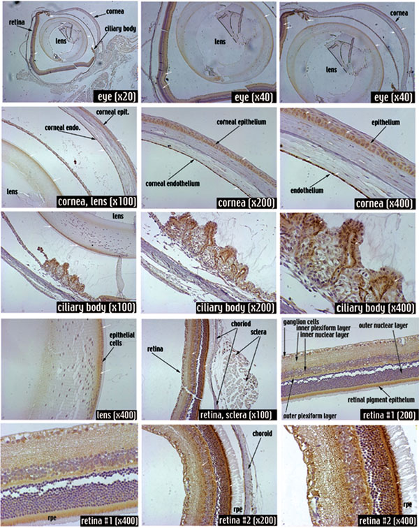
Figure 4. Immunohistochemistry analysis of Chad in mouse ocular tissues
Chad expression was detected in epithelial and endothelial layers of the cornea, the lens, the ciliary body, retinal rod and cone cells, and the plexiform layers. Positive immunostaining appears brown and is highlighted by arrows.