![]() Figure 5 of
Chen, Mol Vis 2003;
9:735-746.
Figure 5 of
Chen, Mol Vis 2003;
9:735-746.
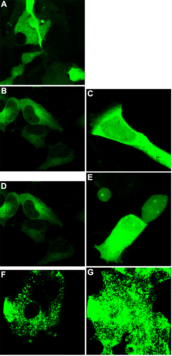
Figure 5. Immunofluorescent analysis of GMP19 and truncations
A: Empty vector pEGFP-N1 transfected T-Rex-293 cells. These cells express free EGFP, which remains soluble in the cytoplasm. B and C: GMP19-25 truncation cell line. The fluorescent signal clearly is associated with the cytoplasm, identical to free EGFP expression alone. D and E: GMP19-36 truncation cell line. Again, the fluorescent signal is found free in the cytoplasm. F and G: GMP19 cell line. Clearly this intact MP19 protein with EGFP fused to the NH2-terminal end associates with the cell membrane, probably with lipid rafts. GMP19-25 and GMP19-36, upon induction with tetracycline express fusion protein that has EGFP at the NH2-terminal end of the polypeptide. The expressed protein remains soluble in the cytoplasm, identical to EGFP alone. GMP19 protein, which has EGFP fused to the NH2-terminal end of MP19, appears to transport to the cell membrane and collect in "islands", much as intact MP19G (Figure 3A,B), which has EGFP fused to the COOH-terminal end of MP19. Analysis conditions are the same as in Figure 3.
