![]() Figure 3 of
Chen, Mol Vis 2003;
9:735-746.
Figure 3 of
Chen, Mol Vis 2003;
9:735-746.
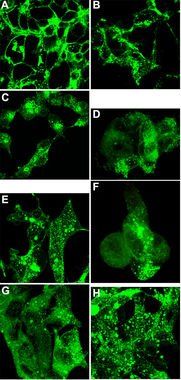
Figure 3. Expression of MP19-EGFP and various truncations of the MP19 molecule
T-REx-293 (293) cells were transfected with either MP19-green-pcDNA4/TO (MP19G) or different truncations of MP19 (MP19-25-green-pcDNA4/TO [MP19-25G], MP19-36-green-pcDNA4/TO [MP19-36G], or MP19-64-green-pcDNA4/TO [MP19-64G]) vectors and stable clones were picked following Zeocin selection. Stable clones were picked and isolated. Each of the cloned expression cell lines was seeded onto glass coverslips and induced with tetracycline when the cells reached the correct cell density. Fluorescent cells were then observed with confocal microscopy as described in [20]. A and B: Confocal microscopy analysis of MP19G expression in 293 cells (EGFP filter set). The image was obtained using a Nikon Optiphot-2 upright microscope with a 100x oil objective. C and D: Confocal microscopy analysis of MP19-25G expression in 293 cells. E and F:Confocal microscopy analysis of MP19-36G expression in 293 cells. G and H:Confocal microscopy analysis of MP19-64G expression in 293 cells. All conditions were the same as those for A and B. All of the truncations appear to be associated with the cell membrane and to sequester in pools within the membrane.